Ascomycota
Ascomycota are commonly know as sac fungi, cup fungi, earth tongues, cramp balls, dung buttons, truffles or moulds.
Most common moulds belonging to the Hyphomycetes are ascomycetes. They may be saprobes, parasites (especially of plants), or lichen forming, mostly terrestrial; cosmopolitan (50 orders, 275 families, 3328 genera, 32,325 spp). Ascomycetes are characterized by septate hyphae with simple pores. Asexual reproduction by conidia. Sexual reproduction by ascospores, typically eight, in an ascus. Asci are often housed in a fruiting body or ascocarp e.g. cleistothecia or perithecia.
Click images below to expand:
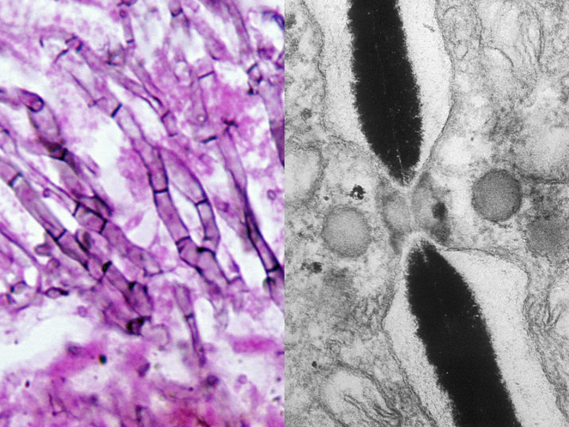


Ascomycetes are characterised by having septate hyphae with simple septal pores showing characteristic Woronin bodies which can plug the pore in the event of injury to the hyphal segment. Asci are often housed in a fruiting body or ascocarp. The above photos show a perithecium (an ostiolate ascocarp) of Gelasinospora. Note the apical ostiole and ascospore discharge through the apical ostiole of a perithecium. Asci typically hold 8 ascospores and which may be single or multi celled.
Species descriptions
-
Aphanoascus flavescens
Aphanoascus fulvescens is a soil-borne keratinolytic ascomycete that occasionally causes dermatomycosis in humans and animals. It has a worldwide distribution and is more commonly found in keratin enriched soils from the degradation of animal hair and skin.
RG-2 organism.

Aphanoascus flavescens culture and cleistothecium.
Morphological description:
Colonies are moderately fast growing, white to tan with the production of numerous spherical, pseudoparenchymatous, buff to light brown cleistothecia (non-ostiolate ascocarps). Asci are subspherical to ellipsoidal and eight-spored. Ascospores light brown, yellowish to pale brown in mass, irregularly reticulate, lens-shaped, 3.5-4.7 x 2.5-3.5 µm. Aphanoascus fulvescens has a Chrysosporium anamorph showing typical pyriform to clavate-shaped conidia with truncated bases, 15-17.5 x 3.7-6 µm, which are formed either intercalary, laterally or terminally.
Aphanoascus fulvescens showing typical pyriform to clavate-shaped conidia with truncated bases.
Molecular identification:
ITS sequencing will differentiate most species. The calmodulin gene may also be useful (Cano et al. 2002, Halliday et al. 2015).Key features:
Keratinolytic, cleistothecia, and a Chrysosporium anamorph.References:
- Cano, J. and Guarro, J. (1990) The genus Aphanoascus. Mycological Research, 94, 355-377.
- Cano, J., Sagues, M., Barrio, E., et al. (2002) Molecular taxonomy of Aphanoascus and description of two new species from soil. Studies in Mycology, 47, 153-164.
- de Hoog, G.S., Guarro, J., Gene, J., et al. (2015) Atlas of Clinical Fungi (Version 4.1.2). Centraalbureau voor Schimmelcultures, Utrecht, The Netherlands.
- Domsch, K.H., Gams, W. and Anderson, T.H. (2007) Compendium of soil fungi. Second Edition, IHW-Verlag, Germany.
- Halliday, C.L., Kidd, S.E., Sorrell, T.C., et al. (2015) Molecular diagnostic methods for invasive fungal disease: the horizon draws nearer? Pathology, 47, 257-269.
- McGinnis, M.R. (1980) Laboratory handbook of medical mycology. Academic Press, New York.
-
Chaetomium spp.
Chaetomium was formerly a large genus with over 250 phenotypic species described. Species were identified by the size and shape of the perithecia, the setae or hairs covering the perithecia, and ascospore morphology.Several species were thermophilic and could grow at temperatures above 37oC. However recent multigene phylogenetic studies have recognised only 44 Chaetomium species, and many others have been reclassified in new genera (Wang et al., 2016, 2022). Molecular identification using the β-tubulin and RPB2 genes is now required to accurately identify species. Chaetomium species are important agents for the decomposition of cellulose waste and plant materials and are only rarely isolated in medical mycology laboratories.
RG-1 organisms.
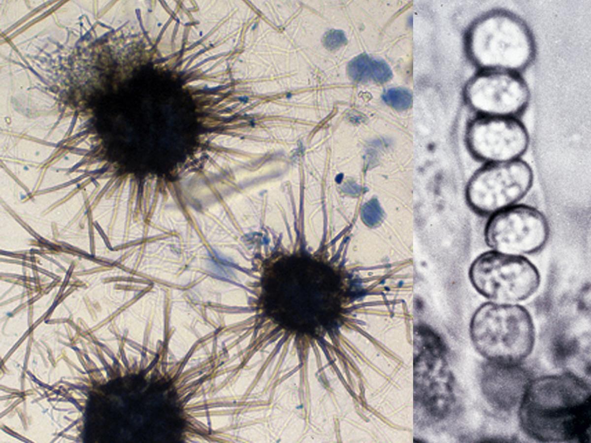
Chaetomium spp. ascocarps (perithecia) with dark coloured terminal hairs (setae), and an ascus with ascospores.
Morphological description:
Chaetomium is a common ascomycetous fungus characterised by the formation of darkly pigmented, globose, ovoid, barrel to flask-shaped, ostiolate ascocarps (perithecia) beset with dark coloured terminal hairs (setae) which are straight, branched or curved. Asci are clavate to cylindrical, typically eight-spored and evanescent. Ascospores are one-celled, darkly pigmented, smooth-walled, of varying shape, mostly ovoid, ellipsoidal or lemon-shaped. Chlamydospores and solitary conidia may also be produced.Molecular identification:
Lee and Hanlin (1999) established the phylogenetic relationships of Chaetomium based on ribosomal DNA sequences. Sequencing of the β-tubulin and RPB2 genes is recommended for routine identification (Wang et al., 2016, 2022).Key features:
Ascomycete producing darkly-pigmented ostiolate perithecia beset with long dark terminal setae.References:
- Ames, L.M. (1963) A monograph of the Chaetomiaceae. U.S. Army Research and Development Serial, 2, 1-125.
- Seth, H.K. (1970) A Monograph of the genus Chaetomium. J. Cramer, Lehre.
- Millner, P.D. (1977) Radial growth responses to temperature by 58 Chaetomium species, and some taxonomic relationships. Mycologia, 69, 492-502.
- Ellis, D.H. (1981) Ascocarp morphology and terminal hair ornamentation in thermophilic Chaetomium species. Mycologia, 73, 755-773.
- Ellis, D.H. and Keane, P.J. (1981) Thermophilic fungi Isolated from some Australian soils. Australian Journal of Botany, 29, 689-704
- von Arx, J.A., Guarro, J. and Figueras, M.J., (1986) The Ascomycete genus Chaetomium. J. Cramer, Berlin.
- Domsch, K.H., Gams, W. and Anderson, T.H. (2007) Compendium of soil fungi. Second Edition, IHW-Verlag, Germany.
- Wang, X.W., Lombard, L., Groenewald, J.Z., et al. (2016) Phylogenetic reassessment of the Chaetomium globosum species complex. Persoonia, 36, 83-133.
- Wang, X.W., Han P.J., Bai, F.Y., et al. (2022) Taxonomy, phylogeny and identification of Chaetomiaceae with emphasis on thermophilic species. Studies in Mycology, 101, 121-243.
Antifungal susceptibility: Chaetomium species (Australian national data); MIC µg/mL. Antifungal No ≤0.016 0.03 0.06 0.125 0.25 0.5 1 2 4 ≥8 AMB 8 5 2 1 ISAV 3 1 1 1 VORI 8 1 3 2 1 1 POSA 8 1 1 4 1 1 ITRA 8 2 5 1