Entomorphthoromycosis
Infections caused by entomophthoraceous fungi:
Zygomycosis due to entomophthoraceous fungi:
This is caused by species of two genera Basidiobolus and Conidiobolus. Infections are chronic, slowly progressive and generally restricted to the subcutaneous tissue in otherwise healthy individuals. Other characteristics that separate these infections from those caused by mucoraceous fungi are a lack of vascular invasion or infarction and the production of a prolific chronic inflammatory response, often with eosinophils and Splendore-Hoeppli phenomena around the hyphae.
Zygomycosis caused by Basidiobolus ranarum.
Zygomycosis caused by Basidiobolus ranarum:
This is a chronic inflammatory or granulomatous disease generally restricted to the subcutaneous tissue of the limbs, chest, back or buttocks, primarily occurring in children and with a predominance in males. Initially, lesions appear as subcutaneous nodules which develop into massive, firm, indurated, painless swellings which are freely movable over the underlying muscle, but are attached to the skin which may become hyperpigmented but not ulcerated.

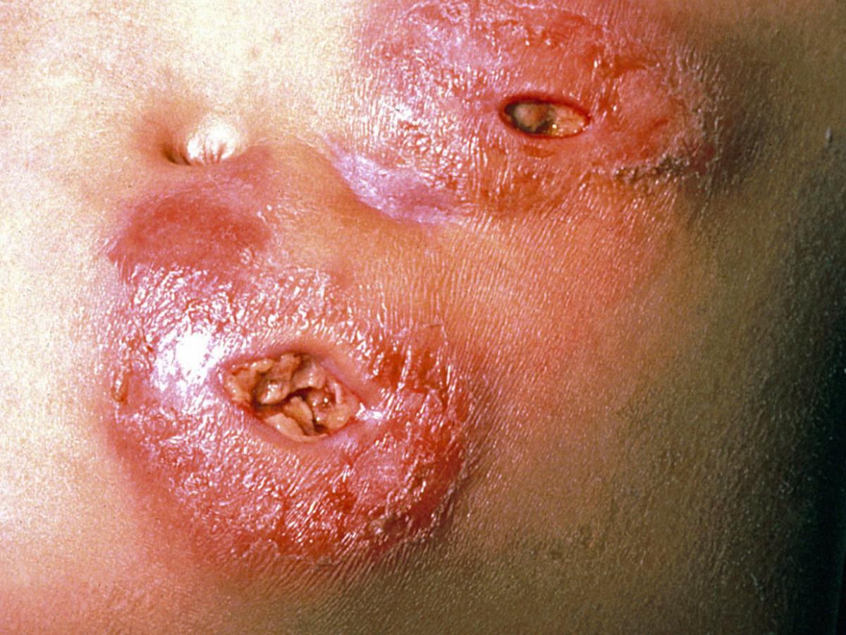
Zygomycosis caused by Conidiobolus (Courtesy of John Rippon, USA).
Zygomycosis caused by Conidiobolus sp.:
This is a chronic inflammatory or granulomatous disease that is typically restricted to the nasal submucosa and characterised by polyps or palpable restricted subcutaneous masses. Clinical variants, including pulmonary and systemic infections have also been described. Human infections occur mainly in adults with a predominance in males (80% of cases). Most cases have been reported from the tropical rain forest areas of central and west and south and central America. Infections usually begin with unilateral involvement of the nasal mucosa. Symptoms include nasal obstruction, drainage and sinus pain. Subcutaneous nodules develop in the nasal and perinasal regions and progressive generalised facial swelling may occur. Infections also occur in horses usually producing extensive nasal polyps and other animals. Conidiobolus coronatus is also a recognised pathogen of termites, other insects and spiders.

H&E stained section of infected tissue showing broad, infrequently septate, hyphae surrounded by an eosinophilic sheath [Splendore-Hoeppli phenomena], typical of zygomycosis caused by Basidiobolus ranarum.
Laboratory Diagnosis:
1. Clinical Material:
Skin biopsy tissue.
2. Direct Microscopy:
Tissue sections should be stained with H&E and GMS. Examine specimens for broad, infrequently septate, thin-walled hyphae, which often show focal bulbous dilations and irregular branching.
Further reading:
Ellis, DH. 2005. Subcutaneous Zygomycetes – Subcutaneous zygomycosis. Chapter 17. In Topley and Wilson’s Microbiology and Microbial Infections: Medical Mycology, 10th edition, Hodder Arnold London pp 347-355.
Kwon-Chung KJ and JE Bennett 1992. Medical Mycology Lea & Febiger.
Rippon JW. 1988. Medical Mycology WB Saunders Co.
Back to Subcutaneous Mycoses Back to Opportunistic Systemic Mycoses