Dimorphic Fungal Pathogens
These are fungal infections of the body caused by fungal pathogens which can overcome the physiological and cellular defences of the normal human host by changing their morphological form.
They are geographically restricted and the primary site of infection is usually pulmonary, following the inhalation of conidia.
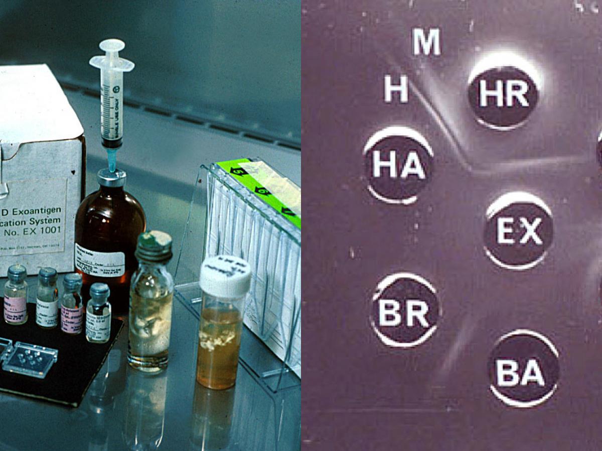
Exoantigen immunodiffusion plate showing positive identification of Histoplasma capsulatum. Note: H and M bands of identification; EX = culture filtrate; H = Histoplasma antibody and antigen, C = Coccidioides antibody and antigen; B = Blastomyces antibody and antigen.
Note: DNA sequencing is now becoming the method of choice to minimise staff exposure to the infectious propagules.
Traditionally, positive identification required conversion of the mould form to the yeast phase by growth at 37C on enriched media, however for laboratory safety, culture identification by either exoantigen test or DNA sequencing is now preferred.
WARNING: RG-3 organisms.
Cultures of these fungi represent a severe biohazard to laboratory personnel and must be handled with extreme caution in an appropriate pathogen handling cabinet.
Species descriptions
-
Blastomyces dermatitidis
Blastomyces contains a number of pathogenic species, most notably Blastomyces dermatitidis and Blastomyces gilchristii, which are morphologically identical but distinguishable by sequence analysis of the ITS region (Brown et al., 2013).
These are associated with soil and decaying organic matter such as leaves and wood in North America. Blastomyces percursus and B. emzantsi have been identified from a culture repository in South Africa (Dukik et al., 2017; Jiang et al., 2018; Maphanga et al., 2020). In addition, Blastomyces helices and B. parvus have been transferred to this genus from Emmonsia (Peterson and Sigler, 1998; Jiang et al., 2018). These are the causative agents of blastomycosis a chronic granulomatous and suppurative disease, having a primary pulmonary stage that is frequently followed by dissemination to other body sites, typically the skin and bone.
WARNING: RG-3 organism.
Cultures of Blastomyces species represent a biohazard to laboratory personnel and must be handled in a Class II Biological Safety Cabinet (BSCII).
Blastomyces dermatitidis culture and microscopy showing one-celled, smooth-walled conidia borne on short lateral to terminal hyphal branches (images provided by Jon Biehle, Dept. of Pathology, Creighton University, Omaha, NE, USA).
Morphological description:
Colonies at 25C have variable morphology and growth rate. They may grow rapidly, producing a fluffy white mycelium or slowly as glabrous, tan, nonsporulating colonies. Growth and sporulation may be enhanced by yeast extract. Most strains become pleomorphic with age. Microscopically, hyaline, ovoid to pyriform, one-celled, smooth-walled conidia (2-10 µm in diameter) of the Chrysosporium type, are borne on short lateral or terminal hyphal branches.Colonies on blood agar at 37C are wrinkled and folded, glabrous and yeast-like. Microscopically, the organism produces the characteristic yeast phase seen in tissue pathology; ie. B. dermatitidis is a dimorphic fungus.
Comment:
In the past, conversion from the mould form to the yeast form was necessary to positively identify this dimorphic pathogen from species of Chrysosporium or Sepedonium. However, culture identification by exoantigen test and/or molecular methods is now preferred to minimise manipulation of the fungus.Key features:
Clinical history, tissue pathology, culture identification by positive exoantigen test and/or by molecular methods.
Tissue section showing large, broad-base, unipolar budding yeast-like cells of Blastomyces dermatitidis.
Histopathology:
Blastomyces dermatitidis tissue sections show large, broad-based, unipolar budding yeast-like cells, which may vary in size from 8-15 µm, with some larger forms up to 30 µm in diameter. Tissue sections need to be stained by Grocott’s methenamine silver method to clearly see the yeast-like cells, which are often difficult to observe in H&E preparations.Molecular diagnostics:
A DNA probe assay (AccuProbe, Gen-Probe, Inc., San Diego, CA) for identification of B. dermatitidis in clinical isolates is available (Scalarone et al. 1992 and Padhye et al. 1994b). However this has limited application as it can be used only with pure cultures of B. dermatitidis (yeast or mould) (Sidamonidze et al. 2012). Several conventional PCR assays have been developed for the identification of B. dermatitidis from clinical specimens (Bialek et al. 2003) and soil (Burgess et al. 2006). Sidamonidze et al. (2012) developed a real-time PCR targeting the BAD1 (formerly known as WI-1) gene for the identification of B. dermatitidis in culture and tissue and Morjaria et al. (2015) used rDNA sequencing for identification from paraffin embedded tissue.Antifungal Susceptibility: B. dermatitidis limited data available (Sugar and Liu 1996, Espinel-Ingroff et al. 2001, Espinel-Ingroff 2003, Gonzales et al. 2005 and Sabatelli et al. 2006); MIC µg/mL.
Antifungal susceptibility testing not recommended.
Antifungal Range MIC90 Antifungal Range MIC90 FLU 0.125-64 4-16 AMB 0.03-1 0.5 ITRA 0.03->16 0.125-2 VORI 0.03-16 0.25 POSA 0.03-2 0.125 CAS 0.5-8 2 References:
- Bialek, R., Cirera, A.C., Herrmann, T., et al. (2003) Nested PCR assays for detection of Blastomyces dermatitidis DNA in paraffin-embedded canine tissue. Journal of Clinical Microbiology, 41, 205-208.
- Brown, E.M., McTaggart, L.R., Zhang, S.X., et al. (2013) Phylogenetic analysis reveals a cryptic species Blastomyces gilchristii, sp. nov. within the human pathogenic fungus Blastomyces dermatitidis. PLoS One, 8, e59237.
- Burgess, J.W., Schwan, W.R. and Volk, T.J. (2006) PCR-based detection of DNA from the human pathogen Blastomyces dermatitidis from natural soil samples. Medical Mycology, 44, 741-748.
- Chandler, F.W., Kaplan, W.and Ajello, L. (1980) A colour atlas and textbook of the histopathology of mycotic diseases. Wolfe Medical, London.
- Dukik, K., Munoz, J.F., Jiang, Y., et al. (2017) Novel taxa of thermally dimorphic systemic pathogens in the Ajellomycetaceae (Onygenales). Mycoses, 60, 296-309.
- Espinel-Ingroff, A., Boyle, K.and Sheehan, D.J. (2001) In vitro antifungal activities of voriconazole and reference agents as determined by NCCLS methods: review of the literature. Mycopathologia, 150, 101-115.
- Espinel-Ingroff, A. (2003) In vitro antifungal activities of anidulafungin and micafungin, licensed agents and the investigational triazole posaconazole as determined by NCCLS methods for 12,052 fungal isolates: review of the literature. Revista Iberoamericana de Micologia, 20, 121-136.
- Gonzalez, G.M., Fothergill, A.W., Sutton, D.A., et al. (2005) In vitro activities of new and established triazoles against opportunistic filamentous and dimorphic fungi. Medical Mycology, 43, 281-284.
- Jiang, Y., Dukik, K., Munoz, J.F., et al. (2018) Phylogeny, ecology and taxonomy of systemic pathogens and their relatives in Ajellomycetaceae (Onygenales): Blastomyces, Emergomyces, Emmonsia, Emmonsiellopsis. Fungal Diversity, 90, 245-291.
- Kaufman, L. and Standard, P.G. (1987) Specific and rapid identification of medically important fungi by exoantigen detection. Annual Review of Microbiology, 41, 209-225.
- Kidd, S., Halliday, C., Ellis, D. (2023) Descriptions of Medical Fungi (4th edition). CABI.
- Maphanga, T. G., Birkhead, M., Munoz, J. F., et al. (2020) Human blastomycosis in South Africa caused by Blastomyces percursus and Blastomyces emzantsi sp. nov., 1967 to 2014. Journal of Clinical Microbiology, 58, e01661-19.
- McGinnis, M.R. (1980) Laboratory handbook of medical mycology. Academic Press, New York.
- Morjaria, S., Otto, C., Moreira, A., et al. (2015) Ribosomal RNA gene sequencing for early diagnosis of Blastomyces dermatitidis infection. International Journal of Infectious Diseases, 37, 122-124.
- Peterson, S.W. and Sigler, L. (1998) Molecular genetic variation in Emmonsia crescens and Emmonsia parva, etiologic agents of adiaspiromycosis, and their phylogenetic relationship to Blastomyces dermatitidis (Ajellomyces dermatitidis) and other systemic fungal pathogens. Journal of Clinical Microbiology, 36, 2918-2925.
- Rippon, J.W. (1988) Medical mycology: the pathogenic fungi and the pathogenic actinomycetes, 3rd edition. W,B. Saunders Co, Philadelphia, USA.
- Sabatelli, F., Patel, R., Mann, P.A., et al. (2006) In vitro activities of posaconazole, fluconazole, itraconazole, voriconazole, and amphotericin B against a large collection of clinically important molds and yeasts. Antimicrobial Agents and Chemotherapy, 50, 2009-2015.
- Sidamonidze, K., Peck, M.K., Perez, M., et al. (2012) Real-time PCR assay for identification of Blastomyces dermatitidis in culture and in tissue. Journal of Clinical Microbiology, 50, 1783-1786.
- Sugar, A.M. and Liu, X.P. (1996) In vitro and in vivo activities of SCH 56592 against Blastomyces dermatitidis. Antimicrobial Agents and Chemotherapy, 40, 1314-1316.
-
Coccidioides immitis/posadasii
Coccidioides immitis has been separated into two distinct species: C. immitis and C. posadasii (Fisher et al. 2002). The two species are morphologically identical and can be distinguished only by genetic analysis and different rates of growth in the presence of high salt concentrations (C. posadasii grows more slowly). C. immitis is geographically limited to California’s San Joaquin Valley region and Mexico, whereas C. posadasii is found in California, Arizona, Texas, Mexico and South America..
WARNING: RG-3 organism.
Cultures of Coccidioides immitis/posadasii represent a severe biohazard to laboratory personnel and must be handled with extreme caution in Class II Biological Safety Cabinet (BSCII). Cultures can grow rapidly (within 48 hours on blood agar) and are often first encountered in routine microbiology laboratories, rather than specialised and contained mycology laboratories. Therefore routine bacteriology staff should be made aware of the risks in handling this organism and steps should be taken to limit these risks.
Coccidioides immitis tissue morphology showing typical endosporulating spherules. Young spherules have a clear centre with peripheral cytoplasm and a prominent thick-wall. Endospores (sporangiospores) are later formed within the spherule by repeated cytoplasmic cleavage. Rupture of the spherule releases endospores into the surrounding tissue where they re-initiate the cycle of spherule development.

Culture of Coccidioides immitis
Morphological description:
Colonies of C. immitis and C. posadasii grown at 25C may initially be moist and glabrous, but rapidly become suede-like to downy, greyish-white with a tan to brown reverse. However, considerable variation in growth rate and culture morphology has been noted. Microscopy shows typical single-celled, hyaline, rectangular to barrel-shaped, alternate arthroconidia, 2.5-4 x 3-6 µm in size, separated from each other by a disjunctor cell.
The closely related non-pathogenic soil fungus, Malbranchea spp. produces morphologically similar arthroconidia, and this organism may be used as a surrogate for laboratory investigations and training. Other soil fungi such as Gymnoascus spp. also produce similar arthroconidia.Comment:
Coccidioides immitis and C. posadasii are dimorphic fungi, existing in living tissue as spherules and endospores, and in soil or culture in a mycelial form. Despite its dimorphism, the ‘spherule phase’ will not be observed using routine laboratory procedures,and inducing this phaseshould not be attempted.
Culture identification by either exoantigen test or DNA sequencing is preferred to minimise exposure to the infectious propagule.
Microscopy shows typical single-celled, hyaline, rectangular to barrel-shaped, alternate arthroconidia, 2.5-4 x 3-6 µm in size, separated from each other by a disjunctor cell.
Key features:
Clinical history, tissue pathology, culture identification by ITS sequence analysis.Molecular identification:
In endemic areas a DNA probe for recognition of the species is commercially available (Padhye et al. 1994b). ITS sequencing is recommended for differentiation of species (Tintelnot et al. 2007, Binnicker et al. 2011).Antifungal susceptibility: Coccidioides immitis (Ramani and Chaturvedi 2007); MIC µg/mL.
Antifungal susceptibility testing not recommended.
No ≤0.03 0.06 0.125 0.25 0.5 1 2 4 8 16 ≥32 AmB 45 7 25 8 4 1 FLU 45 1 4 10 27 3 VORI 45 13 9 15 8 POSA 45 25 13 3 4 ITRA 45 23 22 References:
- Barker, B.M., Litvintseva, A.P., Riquelme, M., et al. (2019) Coccidioides ecology and genomics. Medical Mycology, 57, S21-S29.
- Binnicker, M.J., Popa, A.S., Catania, J., et al. (2010) Meningeal coccidioidomycosis diagnosed by real-time polymerase chain reaction analysis of cerebrospinal fluid. Mycopathologia, 171, 285-289.
- Chandler, F.W., Kaplan, W.and Ajello, L. (1980) A colour atlas and textbook of the histopathology of mycotic diseases. Wolfe Medical, London.
- de Hoog, G.S., Guarro, J., Gene, J., et al. (2015) Atlas of Clinical Fungi (Version 4.1.2). Centraalbureau voor Schimmelcultures, Utrecht, The Netherlands.
- Fisher, M.C., Koenig, G.L., White, T.J., et al. (2002) Molecular and phenotypic description of Coccidioides posadasii sp. nov., previously recognized as the non-California population of Coccidioides immitis. Mycologia, 94, 73-84.
- Kidd, S., Halliday, C., Ellis, D. (2023) Descriptions of Medical Fungi (4th edition). CABI.
- Kirkland, T.N. and Fierer, J. (2018) Coccidioides immitis and posadasii; A review of their biology, genomics, pathogenesis, and host immunity. Virulence, 9, 1426-1435.
- McGinnis, M.R. (1980) Laboratory handbook of medical mycology. Academic Press, New York.
- Ramani R. and Chaturvedi, V. (2007) Antifungal susceptibility profiles of Coccidioides immitis and Coccidioides posadasii from endemic and non-endemic areas. Mycopathologia, 163, 315-319.
- Rippon, J.W. (1988) Medical mycology: the pathogenic fungi and the pathogenic actinomycetes, 3rd edition. W,B. Saunders Co, Philadelphia, USA.
- Stevens, D.A. (1980) Coccidioidomycosis: A text. Springer, New York, NY, USA.
- Sigler, L. and Carmichael, J.W. (1976) Taxonomy of Malbranchea and some other hyphomycetes with arthroconidia. Mycotaxon, 4, 349-488.
- Tintelnot, K., de Hoog, G.S., Antweiler, E., et al. (2007) Taxonomic and diagnostic markers for identification of Coccidioides immitis and Coccidioides posadasii. Medical Mycology, 45, 385-393.
-
Histoplasma capsulatum
WARNING: RG-3 organism.
Cultures of Histoplasma capsulatum represent a severe biohazard to laboratory personnel and must be handled with extreme caution in a Class II Biological Safety Cabinet (BSCII).Histoplasma capsulatum has a worldwide distribution, however the Mississippi-Ohio River Valley in the USA is recognised as a major endemic region. Environmental isolations of the fungus have been made from soil enriched with excreta from chicken, starlings and bats.
Histoplasma capsulatum has recently been subdivided into a number of molecular siblings (Sepulveda et al., 2017), which in part show extensive hybridization (Maxwell et al., 2018) and which may be difficult to distinguish in practice (de Almeida et al., 2020). On a global scale, and particularly in South America, the Histoplasma capsulatum complex shows enormous diversity (Rodrigues et al., 2020a). Patients may be infected by several genotypes (Damasceno et al., 2019)
Histoplasmosis is an intracellular mycotic infection of the reticuloendothelial system caused by the inhalation of the fungus. Approximately 95% of cases of histoplasmosis are inapparent, subclinical or benign. The remaining 5% of cases may develop chronic progressive lung disease, chronic cutaneous or systemic disease or an acute fulminating fatal systemic disease. All stages of this disease may mimic tuberculosis. Sporadic cases have been reported in Australia.

Culture of Histoplasma capsulatum showing mould-like growth at 25C and yeast-like growth at 37C.
Morphological description:
Histoplasma capsulatum exhibits thermal dimorphism growing in living tissue or in culture at 37C as a budding yeast-like fungus and in soil or culture at temperatures below 30C as a mould.Colonies at 25C are slow growing, white or buff-brown, suede-like to cottony with a pale yellow-brown reverse. Other colony types are glabrous or verrucose, and a red pigmented strain has been noted (Rippon, 1988). Microscopic morphology shows the presence of characteristic large, rounded, single-celled, 8-14 µm in diameter, tuberculate macroconidia formed on short, hyaline, undifferentiated conidiophores. Small, round to pyriform microconidia, 2-4 µm in diameter, borne on short branches or directly on the sides of the hyphae may also be present.
Colonies at 37C grown on brain heart infusion (BHI) agar containing blood are smooth, moist, white and yeast-like. Microscopically, numerous small round to oval budding yeast-like cells, 3-4 x 2-3 µm in size are observed.

Large, rounded, single-celled, tuberculate macroconidia and small microconidia of H. capsulatum.
Three varieties of Histoplasma capsulatum are recognised, depending on the clinical disease: var. capsulatum is the common cause of histoplasmosis; var. duboisii is the African type and var. farciminosum causes lymphangitis in horses. Histoplasma isolates may also resemble species of Sepedonium and Chrysosporium. Traditionally, positive identification required conversion of the mould form to the yeast phase by growth at 37C on enriched media, however for laboratory safety, culture identification by either exoantigen test or DNA sequencing is now preferred.
Key features:
Clinical history, tissue morphology, culture morphology and positive exoantigen test or DNA sequencing.Molecular identification:
Eliaset al. (2012) developed a multiplex-PCR for identification from cultures. Scheel et al. (2014) developed a loop-mediated isothermal amplification (LAMP) assay for detection directly in clinical samples which is affordable and useful in resource poor facilities. ITS sequencing may also be used for accurate identification (Estrada-Bárcenas et al. 2014, Irinyi et al. 2015).MALD-ToF MS:
Valero et al. (2018) developed a reference database for the identification of Histoplasma capsulatum.Antifungal Susceptibility: Histoplasma capsulatum limited data available (Espinel-Ingroff 2003, Gonzalez et al. 2005, Sabatelli et al. 2006, Brilhante et al. 2012, Kathuria et al. 2014); MIC µg/mL.
Antifungal susceptibility testing not recommended.
Antifungal Filamentous form Yeast form Range MIC90 Range MIC90 AMB ≤0.03-0.5 0.5 0.03-0.5 0.25 FLU 1-125 16 2-8 8 ITRA ≤0.03-1 0.125 (1) ≤0.03-0.25 0.125 VORI ≤0.03-2 0.25 (1) ≤0.03-0.5 0.5 POSA ≤0.03-2 0.125 (2) 0.03-0.5 0.25 References:
- Brilhantea, R.S.N., Fechinea, M.A.B., Mesquita, J.R.L., et al. (2012) Histoplasmosis in HIV-positive patients in Ceará, Brazil: clinical-laboratory aspects and in vitro antifungal susceptibility of Histoplasma capsulatum isolates. Transactions of the Royal Society of Tropical Medicine and Hygiene, 106, 484-488.
- Chandler, F.W., Kaplan, W.and Ajello, L. (1980) A colour atlas and textbook of the histopathology of mycotic diseases. Wolfe Medical, London.
- Damasceno, L.S., Teixeira, M.M., Barker, B.M., et al. (2019) Novel clinical and dual infection by Histoplasma capsulatum genotypes in HIV patients from Northeastern, Brazil. Science Reports, 9, 11789.
- de Almeida, S.M., Imano, E.C.M., Vicente, V.A., et al. (2020) Primary central nervous system infection by Histoplasma in an immunocompetent adult. Mycopathologia, 185, 331-338.
- de Hoog, G.S., Guarro, J., Gene, J., et al. (2015) Atlas of Clinical Fungi (Version 4.1.2). Centraalbureau voor Schimmelcultures, Utrecht, The Netherlands.
- Elias, N.A., Cuestas, M.L., Sandoval, M., et al. (2012) Rapid Identification of Histoplasma capsulatumdirectly from cultures by multiplex PCR. Mycopathologia, 174, 451-456.
- Espinel-Ingroff, A. (2003) In vitro antifungal activities of anidulafungin and micafungin, licensed agents and the investigational triazole posaconazole as determined by NCCLS methods for 12,052 fungal isolates: review of the literature. Revista Iberoamericana de Micologia, 20, 121-136.
- Estrada-Barcenas, D.A., Vite-Garín, T., Navarro-Barranco, H., et al. (2014) Genetic diversity of Histoplasma and Sporothrix complexes based on sequences of their ITS1-5.8S-ITS2 regions from the BOLD System. Revista Iberoamericana Micologia, 31, 90-94.
- George, R.B. and Penn, R.L. (1986) Histoplasmosis. In Fungal diseases of the Lung. eds Sarosi, G.A. and Davies, S.F. Grune and Stratton Inc.
- Gonzalez, G.M., Fothergill, A.W., Sutton, D.A., et al. (2005) In vitro activities of new and established triazoles against opportunistic filamentous and dimorphic fungi. Medical Mycology, 43, 281-284.
- Irinyi, L., Serena, C., Garcia-Hermoso, D., et al. (2015) International Society of Human and Animal Mycology (ISHAM)-ITS reference DNA barcoding database-the quality controlled standard tool for routine identification of human and animal pathogenic fungi. Medical Mycology, 53, 313-337.
- Kathuria, S., P.K. Singh, J.F. Meis et al. (2014) In vitro antifungal susceptibility profile and correlation of mycelial and yeast forms of molecularly characterized Histoplasma capsulatum strains from India. Antimicrobial Agents and Chemotherapy, 58, 5613-5616.
- Kidd, S., Halliday, C., Ellis, D. (2023) Descriptions of Medical Fungi (4th edition). CABI.
- Maxwell, C.S., Sepulveda, V.E., Turissini, D.A., et al. (2018) Recent admixture between species of the fungal pathogen Histoplasma. Evolution Letters, 2, 210-220.
- McGinnis, M.R. (1980) Laboratory handbook of medical mycology. Academic Press, New York.
- Rippon, J.W. (1988) Medical mycology: the pathogenic fungi and the pathogenic actinomycetes, 3rd edition. W,B. Saunders Co, Philadelphia, USA.
- Rodrigues, A.M., Beale, M.A., Hagen, F., et al. (2020) The global epidemiology of emerging Histoplasmaspecies in recent years. Studies in Mycology, 97:100095.
- Sabatelli, F., Patel, R., Mann, P.A., et al. (2006) In vitro activities of posaconazole, fluconazole, itraconazole, voriconazole, and amphotericin B against a large collection of clinically important molds and yeasts. Antimicrobial Agents and Chemotherapy, 50, 2009-2015.
- Scheel, C.M., Zhou, Y., Theodoro, R.C., et al. (2014) Development of a loop-mediated isothermal amplification method for detection of Histoplasma capsulatum DNA in clinical samples. Journal of Clinical Microbiology, 52, 483-488.
- Sepulveda, V.E., Marquez, R., Turissini, D.A., et al. (2017) Genome sequences reveal cryptic speciation in the human pathogen Histoplasma capsulatum. mBio, 8(6), e01339-17.
- Valero, C., Buitrago, M.J., Gago, S., et. al. (2018) A matrix-assisted laser desorption/ionization time of flight mass spectrometry reference database for the identification of Histoplasma capsulatum. Medical Mycology, 56, 307-314.
-
Paracoccidioides brasiliensis/lutzii
WARNING: RG-3 organism.
Cultures of Paracoccidioides brasiliensis/lutzii represent a biohazard to laboratory personnel and should be handled with extreme caution in a Class II Biological Safety Cabinet (BSCII).Recently P. brasiliensis has been recognised as two species: P. brasiliensis and P. lutzii (Teixeira et al. 2014, Theodoro et al.2012). P. brasiliensis/lutzii is geographically restricted to areas of South and Central America. The two species are morphologically very similar; conidia of P. lutzii are elongated whereas those from P. brasiliensis are pyriform. Molecular confirmation is recommended. A novel, uncultivated strain of P. brasiliensis reported to cause cutaneous lesions in dolphins has been moved recently to a new species, Paracoccidioides cetii (Vilela et al., 2021).
Molecular identification: ITS sequencing is helpful (Imai et al., 2000) however immunodominant antigen Gp43 (GP43) is recommended for barcoding (Vilela et al., 2021).

Multiple, narrow base, budding yeast cells "steering wheels"of Paracoccidioides brasiliensis.
Morphological description:
Colonies grown at 25C are slow growing and variable in morphology. Colonies may be flat, wrinkled and folded, glabrous, suede-like or downy in texture, white to brownish with a tan or brown reverse. Microscopically, a variety of conidia may be seen, including pyriform microconidia, chlamydospores and arthroconidia. However, none of these are characteristic of the species, and most strains may grow for long periods of time without the production of conidia.On blood agar at 37C, the mycelium converts to the yeast phase and colonies are white to tan, moist and glabrous and become wrinkled, folded and heaped. Microscopically, numerous large, 20-60 μm, round, narrow base budding yeast cells are present. Single and multiple budding occurs, the latter are thick-walled cells that form the classical “steering wheel” or “mickey mouse” structures that are diagnostic for this fungus, especially in methenamine silver stained tissue sections.
Key features:
Clinical history, tissue pathology, culture identification with conversion to yeast phase at 37C, however molecular identification is now recommended.Antifungal Susceptibility: Paracoccidioides brasiliensis very limited data (McGinnis et al. 1997). MIC µg/mL.
Antifungal susceptibility testing not recommended.
Antifungal Range Antifungal Range FLU <0.125-64 AMB 0.125-4 ITRA <0.03->1 VORI <0.03-2 References:
- Bagagli, E., Theodoro, R.C., Bosco, S.M., et al. (2008) Paracoccidioides brasiliensis: phylogenetic and ecological aspects. Mycopathologia, 165, 197-207.
- Chandler, F.W., Kaplan, W.and Ajello, L. (1980) A colour atlas and textbook of the histopathology of mycotic diseases. Wolfe Medical, London.
- de Hoog, G.S., Guarro, J., Gene, J., et al. (2015) Atlas of Clinical Fungi (Version 4.1.2). Centraalbureau voor Schimmelcultures, Utrecht, The Netherlands.
- Desjardins, C.A., Champion, M.D., Holder, J.W., et al. (2011) Comparative genomic analysis of human fungal pathogens causing paracoccidioidomycosis. PLOS Genetics, 7, e1002345-e1002345.
- Imai, T., Sano, A., Mikami, Y., et al. (2000) A new PCR primer for the identification of Paracoccidioides brasiliensis on rRNA sequences coding the internal transcribed spacers (ITS) and 5.8S regions. Medical Mycology, 38, 323-326.
- Kidd, S., Halliday, C., Ellis, D. (2023) Descriptions of Medical Fungi (4th edition). CABI.
- McGinnis, M.R. (1980) Laboratory handbook of medical mycology. Academic Press, New York.
- McGinnis, M.R., Pasarell, L., Sutton, D.A., et al. (1997) In vitro evaluation of voriconazole against some clinically important fungi. Antimicrobial Agents and Chemotherapy, 41, 1832-1834.
- Rippon, J.W. (1988) Medical mycology: the pathogenic fungi and the pathogenic actinomycetes, 3rd edition. W,B. Saunders Co, Philadelphia, USA.
- Teixeira, M.M., Theodoro, R.C., Oliveira, F.F., et al. (2014) Paracoccidioides lutzii sp. nov.: biological and clinical implications. Medical Mycology, 52, 19-28.
- Theodoro, R.C., Teixeira, M., Soares Felipe, M.S., et.al. (2012) Genus Paracoccidioides: species recognition and biogeographic aspects. Plos One, 7, e37694.
- Vilela, R., Huebner, M., Vilela, C., et al. (2021) The taxonomy of two uncultivated fungal mammalian pathogens is revealed through phylogeny and population genetic analyses. Scientific Reports, 11, 18119-18119.