Paraphyton
Paraphyton cookei
Synonymy:
Microsporum cookei; Nannizzia cajetana
Based on a recent taxonomic revision of the dermatophytes the genus Paraphyton has been created for three species (de Hoog et al. 2017).
Paraphyton cookei is a geophilic fungus which has been isolated from the hair of small mammals showing no clinical lesions. Infection has been reported in rodents, dogs and rarely in humans. It is not known to invade hair in vivo, but produces hair perforations in vitro. P. cookei has a world-wide distribution.
RG-1 organism.

Culture of Paraphyton cookei.
Morphological description:
Colonies are flat, spreading, buff to pale brown, powdery to suede-like, with a slightly raised and folded centre and some radial grooves. Reverse pigment dark reddish-brown. Numerous large, very thick-walled, echinulate (rough) elliptical macroconidia with predominantly five to six septa but may be from two to eight septa. Occasional spiral hyphae may be seen. Moderate numbers of mainly slender clavate with some pyriform microconidia are present.
Key features:
The macroconidia of L. cookei are quite characteristic and diagnostic; the thick walls and usually larger size of the macroconidia distinguish it from N. gypsea.
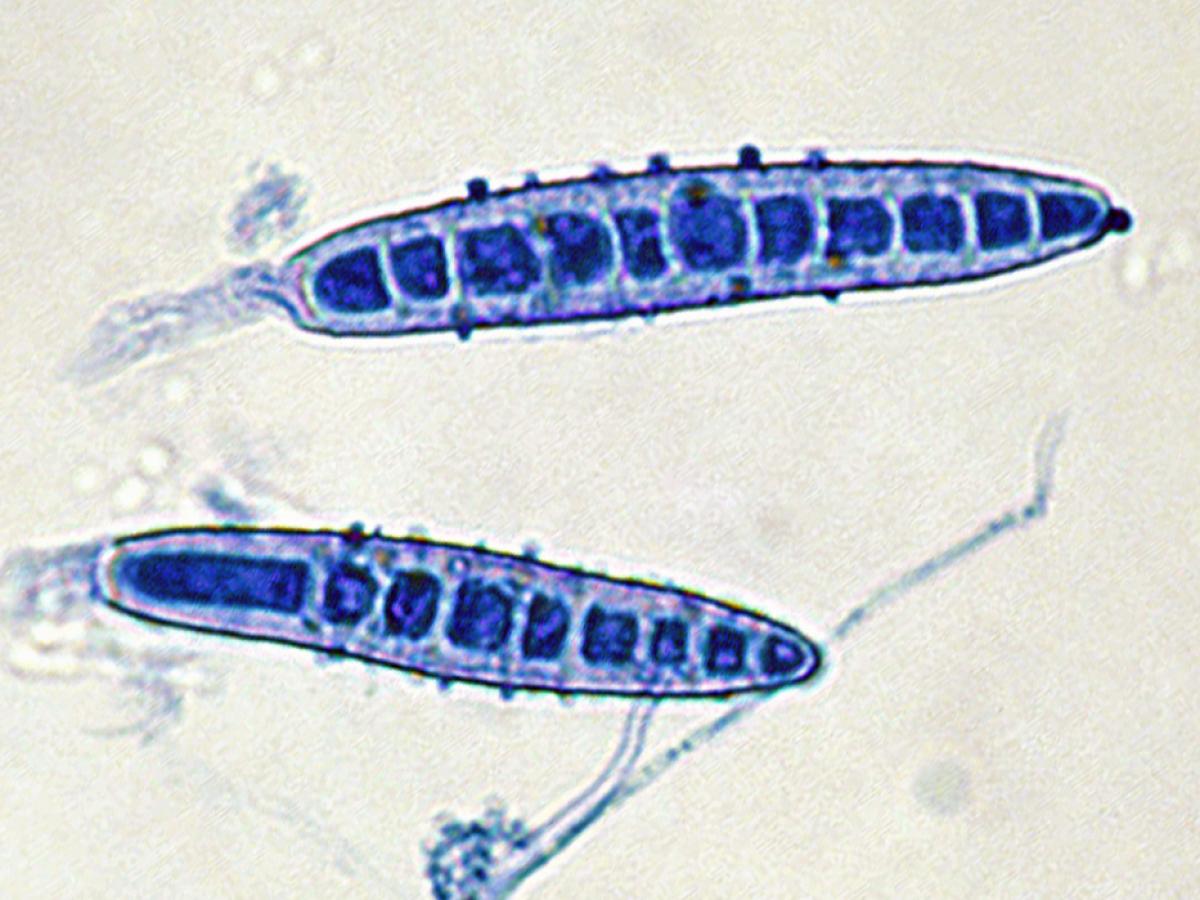
Macroconidia of Paraphyton cookei.
Confirmatory tests:
Vitamin free agar (Trichophyton agar No.1):
Good growth indicating no special nutritional requirements, pinkish-buff-coloured, suede-like colony with a deep magenta red reverse.
Hair perforation test:
Positive.
Molecular identification:
ITS sequencing is recommended.
References:
- Cafarchia, C., Iatta, R., Latrofa, M.S., et al. (2013) Molecular epidemiology, phylogeny and evolution of dermatophytes. Infection, Genetics and Evolution, 20, 336-351.
- Dukik, K., de Hoog, G.S., Stielow, J.B., et al. (2019) Molecular and phenotypic characterization of Nannizzia(Arthrodermataceae). Mycopathologia, 185, 1, 9-35.
- de Hoog, G.S., Guarro, J., Gene, J., et al. (2015) Atlas of Clinical Fungi (Version 4.1.2). Centraalbureau voor Schimmelcultures, Utrecht, The Netherlands.
- de Hoog G.S., Dukik, K., Monod, M., et al. (2017) Towards a noval multilocus phylogenetic taxonomy for dermatophytes. Mycopathologia,182, 5-31.
- Graser, Y., Scott, J. and Summerbell, R. (2008) The new species concept in dermatophytes - a polyphasic approach. Mycopathologia, 166, 239-256.
- Kidd, S., Halliday, C., Ellis, D. (2023) Descriptions of Medical Fungi (4th edition). CABI.
- Rebell, G. and Taplin, D. (1970) Dermatophytes; their recognition and identification. University of Miami Press, FL, USA.
- Rippon, J.W. (1988) Medical mycology: the pathogenic fungi and the pathogenic actinomycetes, 3rd edition. W,B. Saunders Co, Philadelphia, USA.