Curvularia
The genus Curvularia contains about 80 species, which are mostly soil or plant pathogens.
Recent studies have shown that morphological identification does not correlate with molecular identification (Manamgoda et al. 2012; Yanagihara et al. 2010, da Cunha et al. 2013). A phylogenetic analysis of the genera Bipolaris and Curvularia has resulted in a re-alignment of several species. In particular, clinical isolates previously identified as Bipolaris species, notably B. australiensis, B. hawaiiensis and B. spicifera have now been transferred to Curvularia (Manamgoda et al. 2012).
Previously Curvularia lunata was the most frequently reported clinical species, however other species, such as C. americana, C. brachyspora, C. chlamydospora, C. clavata, C. hominis, C. inaequalis, C. muehlenbeckiae, C. pallescens, C. pseudolunata, C. senegalensis and C. verruculosa have now also been reported from clinical cases (Revankar and Sutton, 2010, da Cunha et al. 2013, Madrid et al. 2014).
RG-1 organisms.
Morphological description:
Colonies are fast growing, suede-like to downy, brown to blackish brown with a black reverse. Conidiophores erect, straight to flexuous, septate, often geniculate (producing conidia in sympodial succession) sometimes nodulose. Conidia are ellipsoidal, often curved or lunate, rounded at the ends or sometimes tapering slightly towards the base, pale brown, medium reddish brown to dark brown, 3–10 (usually 3–5) septa, conidial wall smooth to verrucose. Hilum protuberant in some species.
Key features:
Dematiaceous hyphomycete producing sympodial, pale brown, cylindrical or slightly curved phragmoconidia.
Molecular identification:
GPDH (Manamgoda et al. 2012) and/or ITS (da Cunha et al. 2013, Irinyi et al. 2015).
Species descriptions
-
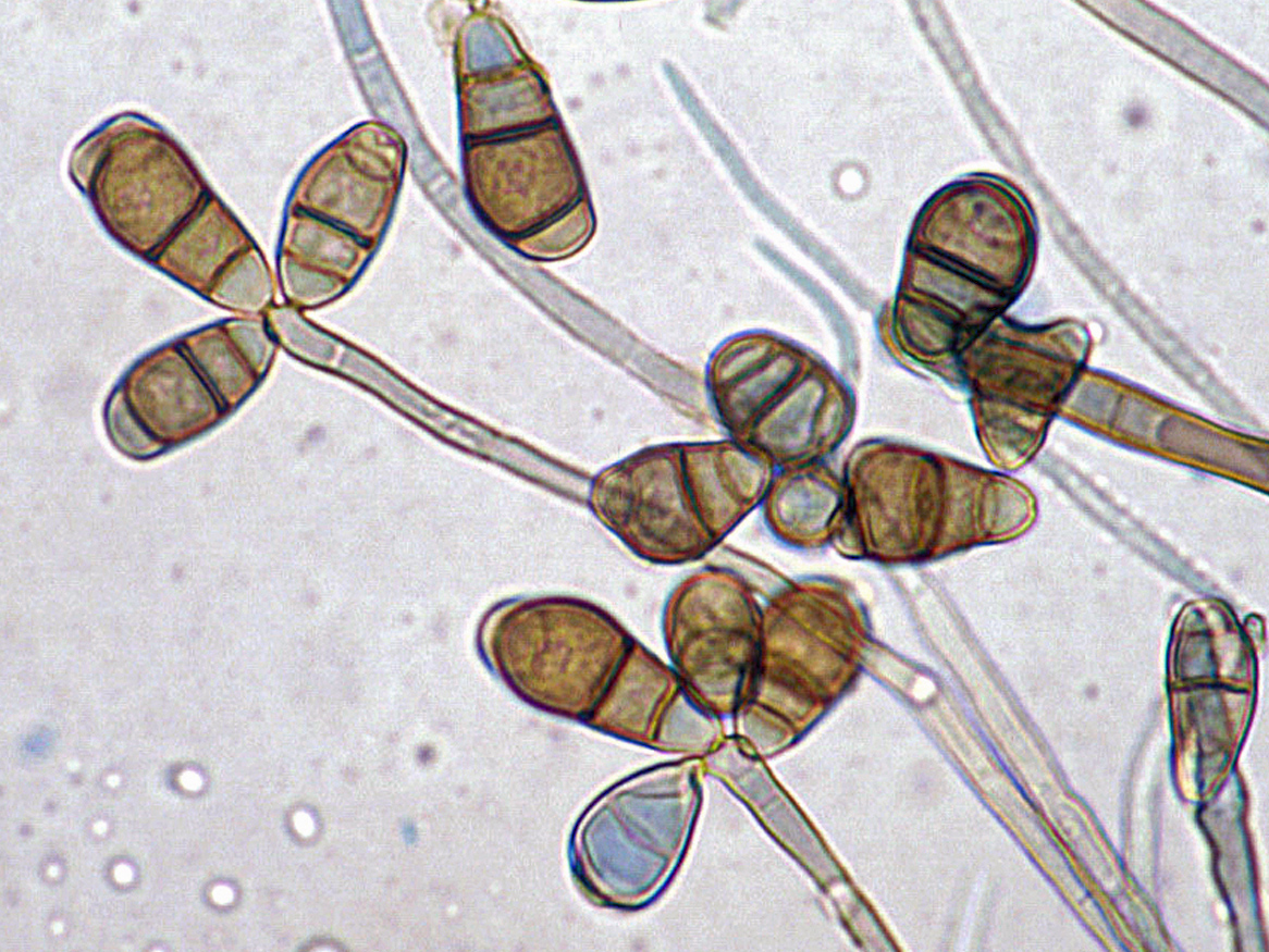
Curvularia australiensis
Curvularia australiensis showing sympodial development of pale brown, fusiform to ellipsoidal, pseudoseptate, poroconidia on a geniculate or zig-zag rachis.
Synonymy: Bipolaris australiensis (M.B. Ellis) Tsuda & Ueyama.
RG-1 organism.
Morphological description:
Colonies are grey to blackish-brown and suede-like in surface texture. Conidiophores are brown, flexuose or geniculate, smooth-walled, up to 150 µm long, mostly 3-7 µm wide. Conidia are straight, rounded at the ends, pale brown to mid reddish-brown, mostly 3-, rarely 4-5-distoseptate, ellipsoidal or oblong, 14-40 × 6-11 µm, smooth-walled to finely roughened.Antifungal susceptibility: Curvularia australiensis (Australian national data); MIC µg/mL. No ≤0.016 0.03 0.06 0.125 0.25 0.5 1 2 4 8 ≥16 AmB 27 1 6 3 7 10 ISAV 4 1 1 2 VORI 27 3 6 7 4 7 POSA 25 1 4 5 6 5 2 2 ITRA 27 1 4 3 13 5 1 -
Curvularia hawaiiensis
Synonymy: Bipolaris hawaiiensis Bugnic.
RG-1 organism.
Morphological description:
Colonies powdery to hairy, black. Conidiophores erect, unbranched, septate, apically flexuose with flat conidial scars on the edges, up to 80 µm long. Conidia smooth- and rather thick-walled, brown, with (3-) 5 (-7) distosepta, cylindrical to cigar-shaped, 18-35 × 6-9 µm.Antifungal susceptibility: Curvularia hawaiiensis (Australian national data); MIC µg/mL. No ≤0.016 0.03 0.06 0.125 0.25 0.5 1 2 4 ≥8 AmB 8 1 4 1 2 ISAV 2 2 VORI 8 2 3 2 1 POSA 6 1 1 1 1 1 1 ITRA 8 1 3 3 1 -
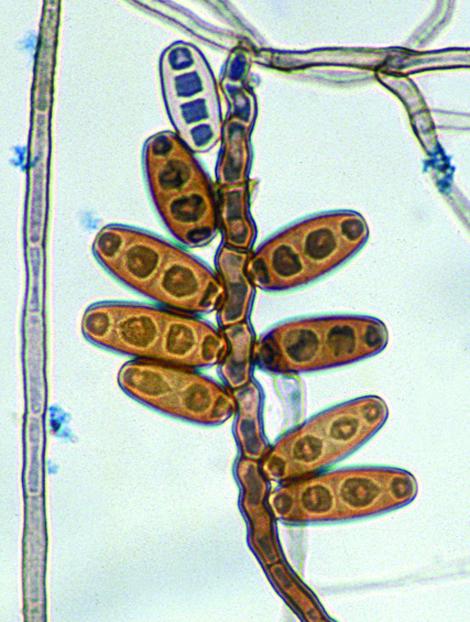
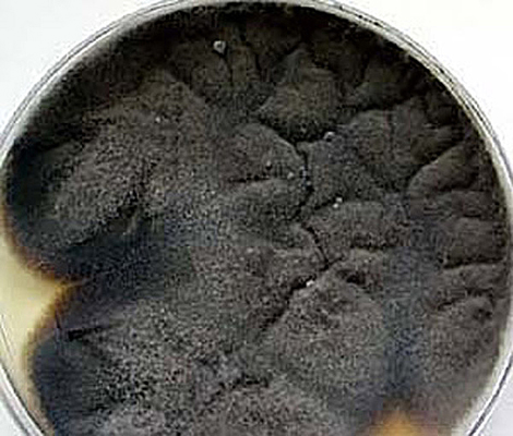
Curvularia lunata
RG-1 organism.
Morphological description:
Colonies black, downy. Conidiophores erect, unbranched, septate, flexuose in the apical part, with flat, dark brown scars. Conidia smooth-walled, olivaceous brown, end cells somewhat paler; conidia obovoidal to broadly clavate, curved at the subterminal cell, 21-31 × 8.5-12.0 µm, 3-septate, the subterminal cell swollen and distinctly larger than the remaining cells.Click images below to enlarge:
Antifungal susceptibility: Curvularia lunata (Australian national data); MIC µg/mL. No ≤0.016 0.03 0.06 0.125 0.25 0.5 1 2 4 8 ≥16 AmB 34 1 5 7 10 8 3 ISAV 4 2 1 1 VORI 34 8 7 9 4 6 POSA 33 2 1 14 10 2 4 ITRA 34 7 16 3 1 1 6 -
Curvularia spicifera
Synonymy: Bipolaris spicifera Bainier.
RG-1 organism.
Morphological description: Colonies appearing glassy with sooty powder of conidia, or hairy if sporulation is poor. Conidiophores erect, unbranched, septate, up to 250 µm long and 4-8 µm wide, regularly zig-zagged in the apical part, with flat, dark brown scars on the edges. Conidia brown, cylindrical with rounded ends, medium brown except for narrow subhyaline spots at the extremites, 20-40 × 9-14 µm, with 3 distosepta.
Antifungal susceptibility: Curvularia spicifera (Australian national data); MIC µg/mL. No ≤0.016 0.03 0.06 0.125 0.25 0.5 1 2 4 ≥8 AmB 3 1 1 1 ISAV 1 1 VORI 3 1 2 POSA 3 1 1 1 ITRA 3 1 2

References:
- da Cunha, K.C., Sutton, D.A., Fothergill, A.W., et al. (2013) In vitro antifungal susceptibility and molecular identity of 99 clinical isolates of the opportunistic fungal genus Curvularia. Diagnostic Microbiology and Infectious Disease, 76, 168-174.
- de Hoog, G.S., Guarro, J., Gene, J., et al. (2015) Atlas of Clinical Fungi (Version 4.1.2). Centraalbureau voor Schimmelcultures, Utrecht, The Netherlands.
- Domsch, K.H., Gams, W. and Anderson, T.H. (2007) Compendium of soil fungi. Second Edition, IHW-Verlag, Germany.
- Ellis, M.B. (1971) Dematiaceous hyphomycetes. Commonwealth Mycological Institute, Kew, Surrey, England.
- Irinyi, L., Serena, C., Garcia-Hermoso, D., et al. (2015) International Society of Human and Animal Mycology (ISHAM)-ITS reference DNA barcoding database-the quality controlled standard tool for routine identification of human and animal pathogenic fungi. Medical Mycology, 53, 313-337.
- Kidd, S., Halliday, C., Ellis, D. (2023) Descriptions of Medical Fungi (4th edition). CABI.
- McGinnis, M.R. (1980) Laboratory handbook of medical mycology. Academic Press, New York.
- Madrid, H., da Cunha, K.C., Gene, J., et al. (2014) Novel Curvularia species from clinical specimens. Persoonia, 33, 48-60.
- Manamgoda, D.S., Cai, L., McKenzie, E.H.C., et al. (2012) A phylogenetic and taxonomic re-evaluation of the Bipolaris - Cochliobolus - Curvularia Complex. Fungal Diversity, 56, 131-144.
- Revankar, S.G. and Sutton, D.A. (2010) Melanized fungi in human disease. Clinical Microbiology Reviews, 23, 884-928.
- Rippon, J.W. (1988) Medical mycology: the pathogenic fungi and the pathogenic actinomycetes, 3rd edition. W,B. Saunders Co, Philadelphia, USA.
- Yanagihara, M., Kawasaki, M., Ishizaki, H., et al. (2010) Tiny keratotic brown lesions on the interdigital web between the toes of a healthy man caused by Curvularia species infection and a review of cutaneous Curvularia infections. Mycoscience, 51, 224-233.