Pseudopithomyces chartarum
The genus Pseudopithomyces was introduced by Ariyawansa et al. (2015) to accommodate several species which had previously been classified in the genus Pithomyces, notably Pithomyces chartarum.
Pseudopithomyces contains 13 species (Ariyawansa et al., 2015) that are commonly found from a very wide range of plant material, also from air, soil, hay, sawn timber and ceiling plaster. Pseudopithomyces chartarum (formerly Pithomyces chartarum) has long been reported as causing facial eczema of sheep. However, recent molecular evidence has identified at least two additional species Pseudopithomyces sacchari (formerlyPithomyces sacchari) and Pseudopithomyces maydicus (formerly Pithomyces maydicus) (da Cunha et al., 2014). Most human isolates are recovered from skin, nail, respiratory and sinus specimens.
RG-1 organism.
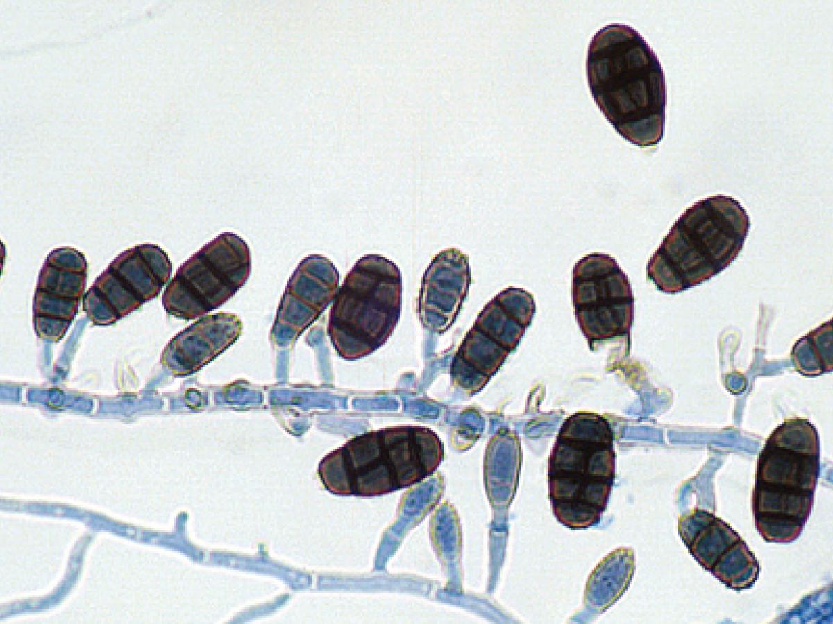
Conidiophore and conidia of Pseudopithomyces chartarum.
Morphological description:
Colonies are fast growing, suede-like to downy and black. Conidiophores are pale olive, smooth or verrucose, 2.5-10 x 2-3.5 µm. Conidiogenous cells integrated, intercalary or terminal, indeterminate, with one to two loci of similar width in the conidiogenous cells. Conidia are muriform, medium to dark brown, echinulate to verrucose, three-(some up to five)-euseptate, slightly constricted at the septa, with one or both median cells divided by longitudinal septa, thick-walled, broadly ellipsoidal, apex obtuse, base truncate and characteristically with part of the conidiogenous cell remaining attached as a small pedicel, 18-29 x 10-17 µm.
Key features:
Dematiaceous hyphomycete with multicelled conidia produced on small peg-like branches of the vegetative hyphae.
Molecular identification:
ITS and D1/D2 sequencing recommended (de Cunha et al. 2014).
| Antifungal susceptibility: Pseudopithomyces spp. (Australian national data); MIC µg/mL. | |||||||||||||
|---|---|---|---|---|---|---|---|---|---|---|---|---|---|
| No | ≤0.03 | 0.06 | 0.125 | 0.25 | 0.5 | 1 | 2 | 4 | 8 | 16 | 32 | ≥64 | |
| AmB | 6 | 1 | 2 | 1 | 1 | 1 | |||||||
| FLU | 6 | 1 | 1 | 1 | 1 | 2 | |||||||
| ISAV | 2 | 1 | 1 | ||||||||||
| VORI | 6 | 1 | 1 | 2 | 2 | ||||||||
| POSA | 6 | 1 | 1 | 4 | |||||||||
| ITRA | 6 | 1 | 1 | 1 | 3 | ||||||||
References:
- Ariyawansa, H.A., Hyde, K.D., Buyck, et al. (2015) Fungal diversity notes 111-252 taxonomic and phylogenetic contributions to fungal taxa, Fungal Diversity, 75, 27-274.
- da Cunha, K.C., Sutton, D.A., Gene, J., et al. (2014) Pithomyces species (Montagnulaceae) from clinical specimens: identification and antifungal susceptibility profiles. Medical Mycology, 52, 748-757.
- de Hoog, G.S., Guarro, J., Gene, J., et al. (2015) Atlas of Clinical Fungi (Version 4.1.2). Centraalbureau voor Schimmelcultures, Utrecht, The Netherlands.
- Domsch, K.H., Gams, W. and Anderson, T.H. (2007) Compendium of soil fungi. Second Edition, IHW-Verlag, Germany.
- Ellis, M.B. (1971) Dematiaceous hyphomycetes. Commonwealth Mycological Institute, Kew, Surrey, England.
- Ellis, M.B. (1976) More dematiaceous hyphomycetes. Commonwealth Mycological Institute, Kew, Surrey, England.
- Kidd, S., Halliday, C., Ellis, D. (2023) Descriptions of Medical Fungi (4th edition). CABI.
- Rippon, J.W. (1988) Medical mycology: the pathogenic fungi and the pathogenic actinomycetes, 3rd edition. W,B. Saunders Co, Philadelphia, USA.