Candidozyma
Two species previously classified in the genus Candida have now been transferred to the genus Candidozyma. Candidozyma auris (formerly Candida auris) and Candidozyma haemuli (formerly Candida haemulonii) (Liu et al. 2024; Borman et al. 2025)
Note. All members of the Candida haemulonii complex have now been transferred to the genus Candidozyma (Candidozyma haemuli, C. pseudohaemuli, and C. duobushaemuli).
-
Candida auris is an emerging multidrug resistant yeast that causes invasive infections, that was first described in 2009 in Japan and has since been reported from numerous countries (Lockhart et al., 2017a,b; Jeffery-Smith et al., 2018; Spivak and Hanson, 2018; Heath et al., 2019; Zhu et al., 2020). Infections and outbreaks caused by C. auris in hospital settings have been rising over the past several years. Difficulty in its identification, multidrug resistance properties, associated high mortality rates, and long-term survival on surfaces in the environment make C. auris particularly problematic in clinical settings (Du et al., 2020). Candida auris is now a notifiable infection under public health regulations in many countries including Australia.

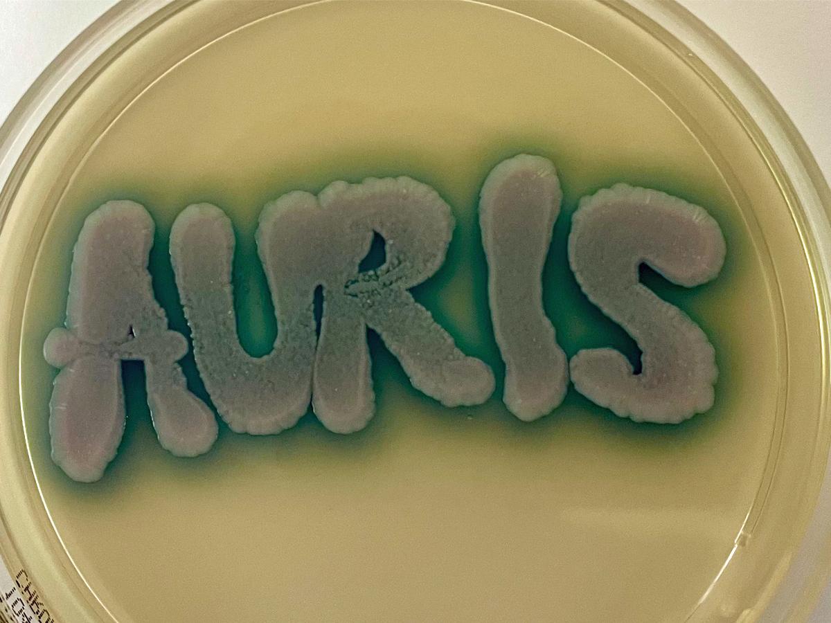
Candida auris grown on CHROMagar™ Candida Plus agar showing characteristic blue halo.
Candida auris may be misidentified as C. haemuloni or other yeast species using conventional phenotypic and biochemical methods (Kathuria et al., 2015; Jeffery-Smith et al., 2018). However, unlike these other Candida species, C. auris grows at 42oC, and this has become a useful differential characteristic (Casadevall et al., 2019). Candida auris is best identified by MALDI-ToF MS or by molecular methods (Du et al., 2020; Zhu et al., 2020).
MALDI-ToF MS: Can accurately differentiate Candida auris from other fungal species; however, accurate identification is dependent on the reference database being used (Kathuria et al., 2015; Jeffery-Smith et al., 2018; Zhu et al., 2020).
Molecular identification: ITS and D1/D2 sequencing provide accurate species identification (Kathuria et al., 2015; Kordalewska et al., 2017; Leach et al., 2018; Du et al., 2020; Zhu et al., 2020).
RG-2 organism.
Culture:
Colonies (SDA) white to cream-coloured smooth, glabrous, yeast-like.Microscopy:
Spherical to subspherical budding blastoconidia, 2-3 x 2-5 µm in size.India ink preparation:
Negative - no capsules present.Dalmau plate culture:
Ovoid budding yeast cells only. No pseudohyphae produced.Physiological tests: (+ Positive, - Negative, v Variable, w Weak, s Slow).
Fermentation:
Glucose + Sucrose + Lactose - Galactose - Maltose - Trehalose w.+ Growth reactions:
Glucose
+
l-Sorbose
-
myo-Inositol
-
Sucrose
+
l-Rhamnose
-
dl-Lactate
-
Raffinose
+
d-Xylose
-
d-Gluconate
-
Melibiose
-
l-Arabinose
-
2-Keto-d-gluconate
-
Galactose
-
d-Arabinose
-
d-Glucosamine
-
Lactose
-
d-Ribose
-
N-Acetyl-d-glucosamine
+
Trehalose
+
Glycerol
-
d-Glucuronate
-
Maltose
+
Erythritol
-
Nitrate
-
Melezitose
+
Ribitol
w,+
Urease
-
Methyl-⍺-d-glucoside
-
Galactitol
+
0.1% Cycloheximide
-
Soluble starch
+
d-Mannitol
+
Growth at 37oC
+
Cellobiose
-
d-Glucitol
+
Growth at 42oC
+,w,s
Key features:
Candida auris is difficult to identify using conventional phenotypic and biochemical methods and has often been misidentified as Candida haemuloni or Saccharomyces cerevisiae. Growth at 42oC on CHROMagar™ Candida produces white, pink, or dark purple colonies (Kumar et al., 2017), however on CHROMagar™ Candida Plus, C. auris colonies display a differential light blue colour with a blue halo (Mulet Bayona et al., 2022). Reliable identification methods are MALDI-ToF MS or ITS and D1/D2 sequencing.
Antifungal susceptibility: Candida auris (Chowdhary et al., 2018; and Australian national data); MIC µg/mL. Antifungal No ≤0.016 0.03 0.06 0.125 0.25 0.5 1 2 4 8 16 32 ≥64 AMB 369 1 27 129 174 22 11 2 FLU 369 5 1 5 2 25 62 269 ISAV 360 44 138 53 38 37 26 11 8 5 VORI 369 21 23 94 91 43 36 28 19 4 10 POSA 369 97 90 85 48 31 8 3 3 2 2 ITRA 369 1 66 79 104 72 28 16 8 1 3 ANID 369 3 28 99 75 112 37 5 1 1 8 MICA 369 26 109 140 61 17 5 1 1 5 4 5FC 369 213 44 5 24 2 11 6 5 9 50
Species descriptions
-
Candida auris is an emerging multidrug resistant yeast that causes invasive infections, that was first described in 2009 in Japan and has since been reported from numerous countries (Lockhart et al., 2017a,b; Jeffery-Smith et al., 2018; Spivak and Hanson, 2018; Heath et al., 2019; Zhu et al., 2020). Infections and outbreaks caused by C. auris in hospital settings have been rising over the past several years. Difficulty in its identification, multidrug resistance properties, associated high mortality rates, and long-term survival on surfaces in the environment make C. auris particularly problematic in clinical settings (Du et al., 2020). Candida auris is now a notifiable infection under public health regulations in many countries including Australia.
Candida auris grown on CHROMagar™ Candida Plus agar showing characteristic blue halo.
Candida auris may be misidentified as C. haemuloni or other yeast species using conventional phenotypic and biochemical methods (Kathuria et al., 2015; Jeffery-Smith et al., 2018). However, unlike these other Candida species, C. auris grows at 42oC, and this has become a useful differential characteristic (Casadevall et al., 2019). Candida auris is best identified by MALDI-ToF MS or by molecular methods (Du et al., 2020; Zhu et al., 2020).
MALDI-ToF MS: Can accurately differentiate Candida auris from other fungal species; however, accurate identification is dependent on the reference database being used (Kathuria et al., 2015; Jeffery-Smith et al., 2018; Zhu et al., 2020).
Molecular identification: ITS and D1/D2 sequencing provide accurate species identification (Kathuria et al., 2015; Kordalewska et al., 2017; Leach et al., 2018; Du et al., 2020; Zhu et al., 2020).
RG-2 organism.
Culture:
Colonies (SDA) white to cream-coloured smooth, glabrous, yeast-like.Microscopy:
Spherical to subspherical budding blastoconidia, 2-3 x 2-5 µm in size.India ink preparation:
Negative - no capsules present.Dalmau plate culture:
Ovoid budding yeast cells only. No pseudohyphae produced.Physiological tests: (+ Positive, - Negative, v Variable, w Weak, s Slow).
Fermentation:
Glucose + Sucrose + Lactose - Galactose - Maltose - Trehalose w.+ Growth reactions:
Glucose
+
l-Sorbose
-
myo-Inositol
-
Sucrose
+
l-Rhamnose
-
dl-Lactate
-
Raffinose
+
d-Xylose
-
d-Gluconate
-
Melibiose
-
l-Arabinose
-
2-Keto-d-gluconate
-
Galactose
-
d-Arabinose
-
d-Glucosamine
-
Lactose
-
d-Ribose
-
N-Acetyl-d-glucosamine
+
Trehalose
+
Glycerol
-
d-Glucuronate
-
Maltose
+
Erythritol
-
Nitrate
-
Melezitose
+
Ribitol
w,+
Urease
-
Methyl-⍺-d-glucoside
-
Galactitol
+
0.1% Cycloheximide
-
Soluble starch
+
d-Mannitol
+
Growth at 37oC
+
Cellobiose
-
d-Glucitol
+
Growth at 42oC
+,w,s
Key features:
Candida auris is difficult to identify using conventional phenotypic and biochemical methods and has often been misidentified as Candida haemuloni or Saccharomyces cerevisiae. Growth at 42oC on CHROMagar™ Candida produces white, pink, or dark purple colonies (Kumar et al., 2017), however on CHROMagar™ Candida Plus, C. auris colonies display a differential light blue colour with a blue halo (Mulet Bayona et al., 2022). Reliable identification methods are MALDI-ToF MS or ITS and D1/D2 sequencing.
Antifungal susceptibility: Candida auris (Chowdhary et al., 2018; and Australian national data); MIC µg/mL. Antifungal No ≤0.016 0.03 0.06 0.125 0.25 0.5 1 2 4 8 16 32 ≥64 AMB 369 1 27 129 174 22 11 2 FLU 369 5 1 5 2 25 62 269 ISAV 360 44 138 53 38 37 26 11 8 5 VORI 369 21 23 94 91 43 36 28 19 4 10 POSA 369 97 90 85 48 31 8 3 3 2 2 ITRA 369 1 66 79 104 72 28 16 8 1 3 ANID 369 3 28 99 75 112 37 5 1 1 8 MICA 369 26 109 140 61 17 5 1 1 5 4 5FC 369 213 44 5 24 2 11 6 5 9 50
-
Candidozyma auris
Synonymy:
Candida aurisCandida auris has recently been placed into the genus Candidozyma based on extensive phylogenetic and phylogenomic analyses (Liu et al. 2024; Borman et al. 2025).
Candidozyma auris is an emerging multidrug resistant yeast that causes invasive infections, that was first described in 2009 in Japan and has since been reported from numerous countries (Lockhart et al., 2017a,b; Jeffery-Smith et al., 2018; Spivak and Hanson, 2018; Heath et al., 2019; Zhu et al., 2020). Infections and outbreaks caused by C. auris in hospital settings have been rising over the past several years. Difficulty in its identification, multidrug resistance properties, associated high mortality rates, and long-term survival on surfaces in the environment make C. auris particularly problematic in clinical settings (Du et al., 2020). Candidozyma auris is now a notifiable infection under public health regulations in many countries including Australia.
Candidozyma auris grown on CHROMagar™ Candida Plus agar showing characteristic blue halo.
Candidozyma auris may be misidentified as Candidozyma haemuli or other yeast species using conventional phenotypic and biochemical methods (Kathuria et al., 2015; Jeffery-Smith et al., 2018). However, unlike these other Candida species, C. auris grows at 42oC, and this has become a useful differential characteristic (Casadevall et al., 2019). Candida auris is best identified by MALDI-ToF MS or by molecular methods (Du et al., 2020; Zhu et al., 2020).
MALDI-ToF MS: Can accurately differentiate Candidozyma auris from other fungal species; however, accurate identification is dependent on the reference database being used (Kathuria et al., 2015; Jeffery-Smith et al., 2018; Zhu et al., 2020).
Molecular identification: ITS and D1/D2 sequencing provide accurate species identification (Kathuria et al., 2015; Kordalewska et al., 2017; Leach et al., 2018; Du et al., 2020; Zhu et al., 2020).
RG-2 organism.
Culture:
Colonies (SDA) white to cream-coloured smooth, glabrous, yeast-like.Microscopy:
Spherical to subspherical budding blastoconidia, 2-3 x 2-5 µm in size.India ink preparation:
Negative - no capsules present.Dalmau plate culture:
Ovoid budding yeast cells only. No pseudohyphae produced.Physiological tests: (+ Positive, - Negative, v Variable, w Weak, s Slow).
Fermentation:
Glucose + Sucrose + Lactose - Galactose - Maltose - Trehalose w.+ Growth reactions:
Glucose
+
l-Sorbose
-
myo-Inositol
-
Sucrose
+
l-Rhamnose
-
dl-Lactate
-
Raffinose
+
d-Xylose
-
d-Gluconate
-
Melibiose
-
l-Arabinose
-
2-Keto-d-gluconate
-
Galactose
-
d-Arabinose
-
d-Glucosamine
-
Lactose
-
d-Ribose
-
N-Acetyl-d-glucosamine
+
Trehalose
+
Glycerol
-
d-Glucuronate
-
Maltose
+
Erythritol
-
Nitrate
-
Melezitose
+
Ribitol
w,+
Urease
-
Methyl-⍺-d-glucoside
-
Galactitol
+
0.1% Cycloheximide
-
Soluble starch
+
d-Mannitol
+
Growth at 37oC
+
Cellobiose
-
d-Glucitol
+
Growth at 42oC
+,w,s
Key features:
Candidozyma auris is difficult to identify using conventional phenotypic and biochemical methods and has often been misidentified as Candidozyma haemuli or Saccharomyces cerevisiae. Growth at 42oC on CHROMagar™ Candida produces white, pink, or dark purple colonies (Kumar et al., 2017), however on CHROMagar™ Candida Plus, C. auris colonies display a differential light blue colour with a blue halo (Mulet Bayona et al., 2022). Reliable identification methods are MALDI-ToF MS or ITS and D1/D2 sequencing.
Antifungal susceptibility: Candidozyma auris (Chowdhary et al., 2018; and Australian national data); MIC µg/mL. Antifungal No ≤0.016 0.03 0.06 0.125 0.25 0.5 1 2 4 8 16 32 ≥64 AMB 369 1 27 129 174 22 11 2 FLU 369 5 1 5 2 25 62 269 ISAV 360 44 138 53 38 37 26 11 8 5 VORI 369 21 23 94 91 43 36 28 19 4 10 POSA 369 97 90 85 48 31 8 3 3 2 2 ITRA 369 1 66 79 104 72 28 16 8 1 3 ANID 369 3 28 99 75 112 37 5 1 1 8 MICA 369 26 109 140 61 17 5 1 1 5 4 5FC 369 213 44 5 24 2 11 6 5 9 50 -
Candidozyma haemuli
Synonymy:
Candida haemuloniiRG-1 organism.
Culture:
Colonies (SDA) white to cream-coloured smooth, glabrous, yeast-like.Microscopy:
Ovoid to globose, budding yeast-like cells or blastoconidia, 2-7 x 2-7 µm. No pseudohyphae produced.India ink preparation:
Negative - no capsules present.Dalmau plate culture:
No pseudohyphae produced.Physiological Tests: + Positive, - Negative, v Variable, w Weak, s Slow, nd No Data Germ Tube - L-Sorbose - L-Arabinose - D-Glucitol + Fermentation Sucrose + D-Arabinose -- 𝝰-M-D-Glucoside - Glucose + Maltose + D-Ribose - D-Gluconate + Galactose - Cellobiose - L-Rhamnose +,w DL-Lactate - Sucrose - Trehalose + D-Glucosamime +,s myo-Inositol - Maltose - Lactose - N-A-D-glucosamine + 2-K-D-Gluconate + Lactose - Melibiose - Glycerol +,s D-Glucuronate - Trehalose +,s Raffinose +,s Erythritol - Nitrate - Assimilation Melezitose +,w Ribitol +,s Urease - Glucose + Soluble Starch - Galactitol - 0.1% Cycloheximide - Galactose +,w D-Xylose - D-Mannitol + Growth at 37C - Key features: Germ tube negative yeast and sugar assimilation pattern. Molecular identification may be required. Candidozyma haemuli has been reported from a few cases of fungaemia but clinical isolations remain rare.
Antifungal susceptibility: Candidozyma haemuli (Australian national data); MIC µg/mL. Antifungal No ≤0.016 0.03 0.06 0.125 0.25 0.5 1 2 4 8 16 32 ≥64 AMB 30 2 10 7 7 1 2 FLU 30 1 3 1 25 VORI 28 1 1 1 1 20 4 POSA 23 1 2 1 1 3 16 ITRA 30 2 1 2 1 24 ANID 21 13 1 3 1 1 2 MICA 21 2 10 3 2 4 CAS 28 1 2 4 6 1 14 5FC 25 2 4 7 6 2 3 1
References
- Borman, A.M., Abdolrasouli, A., and Jhnson, E.M. (2025) Name changes for fungi of medical importance, 2022-2024 Journal of Clinical Microbiology, 63 (11):1-9 10.1128/jcm.02041-24.
- Casadevall, A., Kontoyiannis, D.P. and Robert, V. (2019) On the emergence of Candida auris: climate change, azoles, swamps, and birds. mBio, 10, 01397-19.
- Chowdhary, A., Prakash, A., Sharma, C., et al. (2018) A multicentre study of antifungal susceptibility patterns among 350 Candida auris isolates (2009-17) in India: role of the ERG11 and FKS1 genes in azole and echinocandin resistance. Journal of Antomicrobial Chemotherapy, 73, 891-899.
- Du, H., Bing, J., Hu, T., (2020) Candida auris: Epidemiology, biology, antifungal resistance, and virulence. PLoS Pathology, 16, e1008921.
- Heath, C.H., Dyer, J.R., Pang, S., et al. (2019) Candida auris sternal osteomyelitis in a man from Kenya visiting Australia, 2015. Emerging Infectious Diseases, 25, 192-194.
- Jeffery-Smith, A., Taori, S.K., Schelenz, S., et al. (2018) Candida auris: a review of the literature. Clinical Microbiology Reviews, 31, e00029-17.
- Kathuria, S., Singh, P.K., Sharma, C., et al. (2015) Multidrug-resistant Candida auris misidentified as Candida haemulonii: characterization by matrix-assisted laser desorption ionization- time of flight mass spectrometry and DNA sequencing and its antifungal susceptibility profile variability by Vitek 2, CLSI broth microdilution, and Etest method. Journal of Clinical Microbiology, 53, 1823-1830.
- Kordalewska, M., Zhao, Y., Lockhart, S.R., et al. (2017) Rapid and accurate molecular identification of the emerging multidrug-resistant pathogen Candida auris. Journal of Clinical Microbiology, 55, 2445-2452.
- Kumar, A., Sachu, A., Mohan, K., et al. (2017) Simple low cost differentiation of Candida auris from Candida haemulonii complex using CHROMagar Candida medium supplemented with Pal's medium. Revista Iberoamericana de Micologia, 34,109-111.
- Leach, L., Zhu, Y. and Chaturvedi, S. (2018) Development and validation of a real-time PCR assay for rapid detection of Candida auris from surveillance samples. Journal of Clinical Microbiology, 56, e01223-17.
- Lockhart, S.R., Etienne, K.A., Vallabhaneni, S., et al. (2017a) Simultaneous emergence of multidrug-resistant Candida auris on 3 continents confirmed by whole-genome sequencing and epidemiological analyses. Clinical Infectious Diseases, 64, 134-140.
- Lockhart, S.R., Jackson, B.R., Vallabhaneni, S., et al. (2017b) Thinking beyond the common Candidaspecies: Need for species-level identification of Candida due to the emergence of multidrug-resistant Candida auris. Journal of Clinical Microbiology, 55, 3324-3327.
- Liu F, Hu ZD, Zhao XM, Zhao WN, Feng ZX, Yurkov A, Alwasel S, Boekhout T, Bensch K, Hui FL, Bai FY, Wang QM. (2024) Phylogenomic analysis of the Candida auris-Candida haemuli clade and related taxa in the Metschnikowiaceae, and proposal of thirteen new genera, fifty-five new combinations and nine new species. Persoonia 52:22-43. https://doi.org/10.3767/persoonia.2024.52.02
- Mulet Bayona, J.V., Salvador Garcia, C., Tormo Palop, N., et al. (2022) Candida Plus for detection of Candida auris and other Candida species from surveillance and environmental samples: A multicenter study. Journal of Fungi, 8, 281.
- Spivak, E.S. and Hanson, K.E. (2018) Candida auris: an emerging fungal pathogen. Journal of Clinical Microbiology, 56, e01588-17.
- Zhu, Y., O’Brien, B., Leach, L., et al. (2020) Laboratory analysis of an outbreak of Candida auris in New York from 2016 to 2018: Impact and lessons learned. Journal of Clinical Microbiology, 58, e01503-19.