Basidiobolus ranarum
Synonyms:
Basidiobolus meristosporus; Basidiobolus haptosporus; Basidiobolus heterosporus.
Basidiobolus ranarum is commonly present in decaying fruit and vegetable matter, and as a commensal in the intestinal tract of frogs, toads and lizards.
It has been reported from tropical regions of Africa and Asia including India, Indonesia and Australia. B. ranarum is a known cause of subcutaneous and gastrointestinal entomophthoromycosis often as a result of traumatic inoculation or a barrier break (Ellis, 2005; Vilela and Mendoza, 2018).
RG-2 organism.
Morphological description:
Colonies are moderately fast growing at 30C, flat, yellowish-grey to creamy-grey, glabrous, becoming radially folded and covered by a fine, powdery, white surface mycelium. Satellite colonies are often formed by germinating conidia ejected from the primary colony. Microscopic examination usually shows the presence of large vegetative hyphae (8-20 µm in diameter) forming numerous round (20-50 µm in diameter), smooth, thick-walled zygospores that have two closely appressed beak-like appendages. The production of “beaked” zygospores is characteristic of the genus. Two types of asexual conidia are formed, although isolates often lose their ability to sporulate with subculture. Special media incorporating glucosamine hydrochloride and casein hydrolsate may be needed to stimulate sporulation (Shipton and Zahari, 1987). Primary conidia are globose, one-celled, solitary and are forcibly discharged from a sporophore. The sporophore has a distinct swollen area just below the conidium that actively participates in the discharge of the conidium. Secondary (replicative) conidia are clavate, one-celled and are passively released from a sporophore. These sporophores are not swollen at their bases. The apex of the passively released spore has a knob-like adhesive tip. These spores may function as sporangia, producing several sporangiospores.
Click images below to expand:
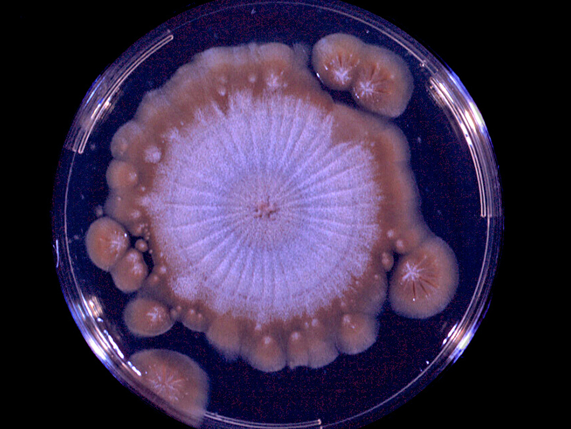

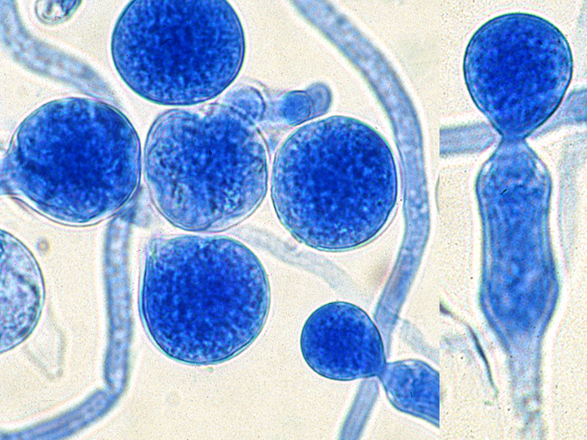
Images of Basidiololus ranarum showing a culture displaying satellite colonies that are often formed by germinating conidia ejected from the primary colony, "beaked" zygospores, conidia and a sporophore with a distinct swollen area just below the conidium.
Molecular identification: ITS sequencing is useful for identification of most clinical isolates. In the past, pathogenic isolates of Basidiobolus were identified as either B. ranarum, B. meristosporus or B. haptosporus,until all three species were found to be identical by rDNA RFLP analysis (Kwon-Chung and Bennett, 1992). However, recent sequence data may suggest the need to re-establish several species (Bshabshe et al., 2020; Al-Hatmi et al., 2021). Many sequences in GenBank are misidentified, so comparison to sequences from Type cultures is important.
Key features: The genus Basidiobolus is distinguished primarily by the morphology and development of forcibly discharged conidia and zygospores, both of which must be observed for reliable identification to genus and species level (King 1983).
References:
- Al-Hatmi ,A.M.S, Balkhair, A., Al-Busaidi, I., et al. (2021) Basidiobolus omanensis sp. nov. causing angioinvasive abdominal Basidiobolomycosis. Journal of Fungi, 7(8), 653.
- Bshabshe A.A., Joseph, M.R.P., Hakami, A.M.A., et al. (2020) Basidiobolus haptosporus-like fungus as a causal agent of gastrointestinal basidiobolomycosis. Medical Mycology, 58, 264-267.
- Davis, S.R., Ellis, D.H., Goldwater, P., et al. (1994) First human culture-proven Australian case of entomophthoromycosis caused by Basidiobolus ranarum. Journal Medical and Veterinary Mycology, 32, 225-230.
- Dworzack, D.L., Pollock, A.S., Hodges, G.R., et al. (1978) Zygomycosis of the maxillary sinus and palate caused by Basidiobolus haptosporus. Archives of Internal Medicine, 138, 1274-1276.
- Ellis, D.H. (2005) Subcutaneous Zygomycetes - Entomophthoromycosis., in Merz, W.G. and Hay, R.J. (eds), Topley and Wilson's Microbiology and Microbial Infections: Medical Mycology, 10 edition. London: Hodder Arnold, 347-355.
- Greer, D.L. and Friedman, L. (1966) Studies on the genus Basidiobolus with reclassification of the species pathogenic for man. Sabouraudia, 4, 231-241.
- Hung, T.-Y., Taylor, B., Lim, A., et al. (2020) Skin and soft tissue infection caused by Basidiobolus spp. in Australia. IDCases, 20, e00731-e00731.
- Kidd, S., Halliday, C., Ellis, D. (2023) Descriptions of Medical Fungi (4th edition). CABI.
- King, D.S. (1983) Entomophthorales. In: Howard D.H. (ed.) Fungi Pathogenic for Humans and Animals. Part A Biology. Marcel Dekker Inc. NY, USA, pp. 61-73.
- Kwon-Chung, K.J. and Bennett, J.W. (1992) Medical Mycology. Lea & Febiger, Philadelphia, 861pp.
- Rippon, J.W. (1988) Medical mycology: the pathogenic fungi and the pathogenic actinomycetes, 3rd edition. W,B. Saunders Co, Philadelphia, USA.
- Shaikh, N., Hussain, K.A., Petraitiene, R., et al. (2016) Entomophthoramycosis: a neglected tropical mycosis. Clinical Microbiology and Infection, 22, 688-694.
- Shipton, W.A. and P. Zahari. 1987. Sporulation media for Basidiobolus species. Journal of Medical and Veterinary Mycology, 25, 323-327.
- Strinivasan, M.C. and Thirumalachar, M.J. (1965) Basidiobolus species pathogenic for man. Sabouraudia, 4, 32-34.
- Vikram, H.R., Smilack, J.D., Leighton, J.A., et al. (2012) Emergence of gastrointestinal basidiobolomycosis in the United States, with a review of worldwide cases. Clinical Infectious Diseases, 54, 1685-1691.
- Vilela, R. and Mendoza, L. (2018) Human pathogenic entomophthorales. Clinical Microbiology Reviews, 31, e00014-18.