Saksenaea complex
The genus Saksenaea is characterised by the formation of flask-shaped sporangia with columellae and simple, darkly pigmented rhizoids. It is an emerging human pathogen (Holland, 1997) that is most often associated with cutaneous or subcutaneous lesions after trauma.
Until recently, Saksenaea vasiformis was the only known species with a worldwide distribution in association with soil. However, recent phylogenetic studies have identified additional species, namely, S. erythrospora, S. dorisiae, S. loutrophoriformis, S. oblongispora and S. trapezispora (Alvarez et al., 2010b; Crous et al., 2016, 2017; Labuda et al., 2019). So far, all species (except S. dorisiae) have been isolated from clinical samples.
Saksenaea vasiformis
RG-2 organism.

Sporangium of Saksenaea vasiformis.
Morphological description:
Colonies are fast growing, downy, white with no reverse pigment, and made up of broad, non-septate hyphae typical of a mucormycetous fungus. Sporangia are typically flask-shaped with a distinct spherical venter and long-neck, arising singly or in pairs from dichotomously branched, darkly pigmented rhizoids. Collumellae are prominent and dome-shaped. Sporangiospores are small, oblong, 1-2 x 3-4 µm, and are discharged through the neck following the dissolution of an apical mucilaginous plug.
Key features:
Mucorales, unique flask-shaped sporangia, failure to sporulate on primary isolation media.
Molecular identification:
ITS sequencing is required for differentiation of species within the complex, and may be necessary to achieve identification in a timely manner (Alvarez et al. 2010b, Walther et al. 2012, Halliday et al. 2015).

Culture of Saksenaea vasiformis.
Agar block method to induce sporulation of Saksenaea vasiformis. A small block of agar is cut from a well established culture grown on PDA and is placed in the centre of petri dish containing 1% agar in distilled water. After 21 days at 26°C look for sporangium formation at the periphery of the petri dish.
Comment:
Laboratory identification of this fungus may be difficult or delayed because of the mould’s failure to sporulate on primary isolation media or on subsequent subculture onto potato dextrose agar. Sporulation may be stimulated by using the agar block method described by Ellis and Ajello (1982), Ellis and Kaminski (1985) and Padhye and Ajello (1988), although this may still take a period of days to weeks. Failure to sporulate prohibits antifungal susceptibility testing.
| Antifungal | Range | MIC90 | Antifungal | Range | MIC90 |
|---|---|---|---|---|---|
| AMB | 0.125-2 | 2 | VORI | 0.5-4 | 4 |
| POSA | 0.016-0.03 | 0.03 | POSA | 0.016-0.25 | 0.25 |
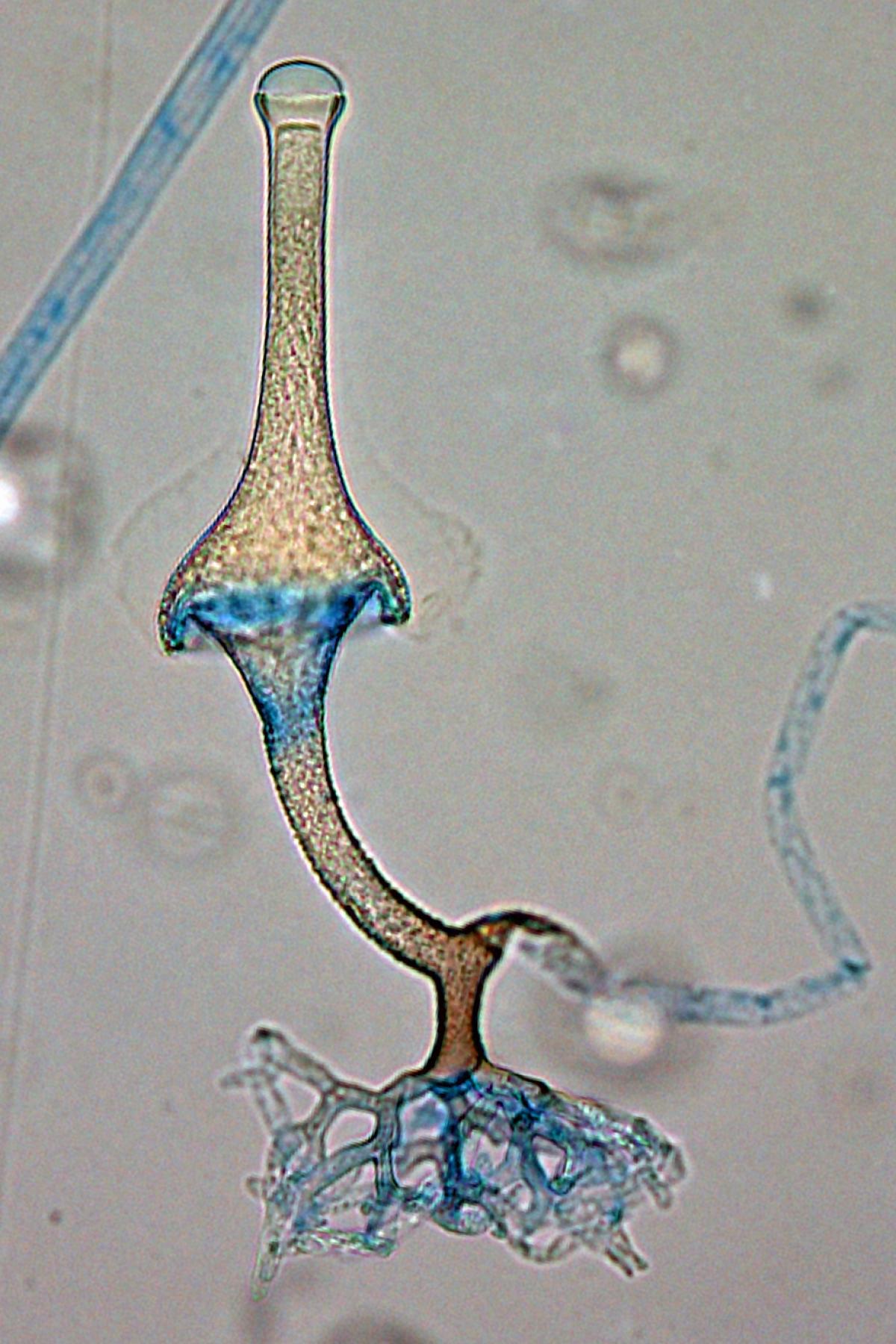
Saksenaea vasiformis sporangium showing distinctive rhizoids.
References:
- Ajello, L., Dean, D.F. and Irwin, R.S. (1976) The zygomycete Saksenaea vasiformis as a pathogen of humans with a critical review of the aetiology of zygomycosis. Mycologia, 68, 52-62.
- Alvarez, E., Sutton, D.A., Cano, J., et al. (2009) Spectrum of zygomycete species identified in clinically significant specimens in the United States. Journal of Clinical Microbiology, 47, 1650-1656.
- Crous, P., Wingfield, M., Burgess, T., et al. (2016) Fungal Planet description sheets: 469-557, Persoonia, 37, 218-403.
- Crous, P., Wingfield, M., Burgess, T., et al. (2017) Fungal Planet description sheets: 558-624, Persoonia, 38, 240-384.
- Davidson, N., Campbell, K., Foroughi, F. et al. (2020) Disseminated Saksenaea infection in an immunocompromised host associated with a good clinical outcome: a case report and review of the literature. BMC Infectious Diseases, 20, 05459-9.
- de Hoog, G.S., Guarro, J., J. Gene, J., et al. (2020) Atlas of clinical fungi. 4th edition. Foundation Atlas of Clinical Fungi https://webshop.atlasclinicalfungi.org.
- Ellis, D.H. (2005) Systemic Zygomycetes - Mucormycosis., in Merz, W.G. and Hay, R.J. (eds), Topley and Wilson’s Microbiology and Microbial Infections: Medical Mycology 10 edition. London: Hodder Arnold, 659-686.
- Ellis, D.H. and Kaminski, G.W. (1985) Laboratory identification of Saksenaea vasiformis: a rare cause of zygomycosis in Australia. Sabouraudia, 23, 137-140.
- Ellis, J.J. and Ajello, L. (1982) An Unusual Source for Apophysomyces elegans and a method for stimulating sporulation of Saksenaea vasiformis. Mycologia, 74, 144-145.
- Ellis, J.J. and Hesseltine, C.W. (1966) Two new families of Mucorales. Mycologia, 66, 87-95.
- Goldschmied-Reouven, A., Shvoron, A., Topaz, M. and Block C. (1989) Saksenaea vasiformis infection in a burn wound. Journal of Medical and Veterinary Mycology, 27, 427-429.
- Halliday, C.L., Kidd, S.E., Sorrell, T.C., et al. (2015) Molecular diagnostic methods for invasive fungal disease: the horizon draws nearer? Pathology, 47, 257-269.
- Holland, J. (1997) Emerging zygomycosis of humans: Saksenaea vasiformis and Apophysomyces elegans. Current Topics in Medical Mycology, 8, 27-34.
- Kidd, S., Halliday, C., Ellis, D. (2023) Descriptions of Medical Fungi (4th edition). CABI.
- Labuda, R., Bernreiter, A., Hochenauer, D., et al. (2019) Saksenaea dorisiae sp. nov., a new opportunistic pathogenic fungus from Europe, International Journal of Microbiology, 2019, 6253829.
- Padhye, A.A. and Ajello, L. (1988) Simple method of inducing sporulation by Apophysomyces elegans and Saksenaea vasiformis. Journal of Clinical Microbiology, 26, 1861-1863.
- Padhye, A.A., Koshi, G., Anandi, V., et. al. (1988) First case of subcutaneous zygomycosis caused by Saksenaea vasiformis in India. Diagnostic Microbiology and Infectious Disease, 9, 69-77.
- Pritchard, R.C., Muir, D.B., Archer, K.H. et. al. (1986) Subcutaneous zygomycosis due to Saksenaea vasiformis in an infant. Medical Journal of Australia, 145, 630-631.
- Saksena, S.B. (1953) A new genus of Mucorales. Mycologia, 45, 426-436.
- Sun, Q.N., Fothergill, A.W., McCarthy, D.I., et. al. (2002) In vivo activities of posaconazole, itraconazole, voriconazole, amphotericin B, and fluconazole against 37 clinical isolates of zygomycetes. Antimicrobial Agents Chemotherapy, 46, 1581-1582.
- Walther, G., Wagner, L. and Kurzai, O. (2019) Updates on the taxonomy of Mucorales with an emphasis on clinically important taxa. Journal of Fungi, 5, 106.